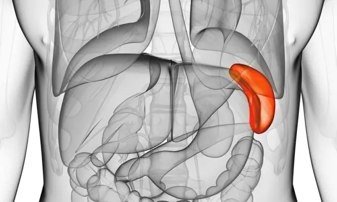
An organ refers to a larger and important part of an organism, made up of tissues that perform similar functions. And one of the major organs in vertebrates, including humans, is the spleen.
It is a large ductless vascular gland; an adult spleen measures a size of about 4 x 7 x 11 cm, weighs around 7 oz. (about 150g-200g).
It is found in the upper left abdomen close to the stomach’s fundus, just below the diaphragm between the 9th and 11th ribs.
Structure of the Spleen
External Structure
The spleen comprises soft tissues, and these tissues are filled with blood trickles (fragile streams of blood) that flow continuously through the parenchyma and clear off old and deformed Red blood cells and White blood cells.
However, the spleen is enveloped by a rigid fibrous capsule structure made up of connective tissue called the tunica fibrosa.
This embedment results in the spleen’s relatively constant “bean shape.” A trabecula runs through the inside of the spleen from the fibrous capsule, forming a supporting framework that divides the spleen into segments.
The spleen has three borders: a superior, an intermediate, and an inferior border. The superior border is notched by the anterior end. The intermediate border of the spleen is directed towards the right.
The inferior border is the rounded part of the spleen. The spleen has two surfaces: the convex diaphragmatic surface, which has a mutual border with the diaphragm and rests on the left kidney’s upper pole.
The second surface is the concave visceral surface that overlays the bowel (stomach) and contains the splenic hilum, where all vessels enter and exit the spleen.
Internal Structure
Internally, the tissues of the spleen divide into two significant units: white pulp and red pulp. The former is composed of lymphatic tissue surrounding a central arteriole, and it mainly contains white blood cells responsible for the initiation of the adaptive immune system response.
The white pulp has an innermost area, the germinal center that contains B-cells, while the marginal zone surrounding the white pulp contains T-cells.
The marginal zone is surrounded by PALS (a periarteriolar lymphoid sheath), which also contain T-cells. Throughout the spleen, the white pulp is surrounded by the red pulp.
The red pulp consists of splenic cords (a.k.a Cords of Billroth or red pulp cords) and a large volume of venous sinuses, pouches, or cavities the tissue made the branching network of veins into the spleen.
This venous network gives the spleen its characteristic red appearance under a microscope. Splenic cords of the red pulp provide the organ structure through reticulin and fibrils, an extensive reservoir of monocytes to aid in wound healing.
Splenic cords also lead to splenic sinuses where macrophages respond to antigens and filter abnormal or old erythrocytes (red blood cells) out of blood flow.
As earlier stated, a thin framework of tough connective tissue known as the tunica fibrosa covers the spleen from which trabeculae arise.
These trabeculae are fibrous bands transporting blood vessels to and from the splenic pulp.
The Spleen and Adjacent Organs
The visceral surface of the spleen borders on various other visceral organs. They include:
Stomach
When the bowel is full, it pushes the spleen moves into a more vertical position. An important radiological visible feature in a plain film of the abdomen is moving the gastric bubble away from the spleen and towards the midline.
These movements may sign of subcapsular hematoma of the spleen or perisplenic hemorrhage such as mononucleosis-induced splenic rupture.
Colon (colic surface)
When an individual is experiencing potent flatulence due to unfavorable ingested food, the spleen moves into a more horizontal position.
Because of the close relationship between the colon and spleen, the left colic flexure is also called the splenic flexure.
Diaphragm (diaphragmatic surface)
The spleen rests very close to the diaphragm and therefore moves alongside the respiratory movement of the diaphragm.
Pancreas
The splenic artery and vein join with the pancreas’ tail, and they abut on the splenic hilum.
Renal Surface or Anterior surface of the Left Kidney
The splenorenal recess lay between the left kidney and the spleen. This recess is an anatomic space where sonography can swiftly detect any free fluid or blood accumulated in the recess from trauma or other pathological events.
Functions of the Spleen
The spleen is one of the secondary lymphatic organs like the thymus. It is the largest lymphatic organ in an adult human.
The spleen is medically referred to as the Central Building (Organ) of the immune system. Also, it filters the blood and rids it of all old and damaged red blood cells (RBCs).
In a macroscopic transected view of the spleen, the parenchyma division into white and red areas, the white and red pulp becomes apparent.
The different coloration of both areas is due to the composition of the various tissues.
During fetal formation and development, the spleen is partly responsible for hemoglobin synthesis, from the 10th to the 25th week of gestation.
After birth, the principal functions of the spleen become different, they include:
- Filtration
- Prevention of infection
- Iron metabolism
- Erythrocytes (Red Blood Cells) storage
- Platelet storage
Filtration of red blood cells and platelets occurs via splenic cords in the red pulp. Flexible younger erythrocytes pass through the epithelial cells of the cords and continue through blood flow.
On the other hand, the older, more giant, and deformed red blood cells are trapped by the splenic cords and phagocytosed by macrophages found in the reticulum and sinus endothelium.
Also, macrophages in the red pulp’s splenic cords are specialized in recycling iron from the breakdown of senescent and damaged erythrocytes.
Macrophages can either store the ingested iron in their cytoplasm or export it via ferritin into the bloodstream.
The spleen does not only breakdown red blood cells as its primary role; it also can play a role in hematopoiesis, the production of all the cellular components of blood and blood plasma.
This role is not a primary function of the spleen in pathologic conditions, such as beta-thalassemia; extramedullary hematopoiesis may be required to assist the bone marrow compensates for the hemolysis taking place.
Prevention of infection occurs by two major roles of the spleen: phagocytic filtration of the blood and opsonizing antibodies’ production.
In addition to supervising red blood cells’ flow, macrophages also monitor microorganisms flowing through the splenic cords, and the macrophages will ingest any foreign body.
Furthermore, in the white pulp’s follicle, infectious antigens and blood-borne microbes are presented by antigen-presenting cells.
This process activates the T-cells and B-cells, which eventually leads to opsonizing antibody production. After the opsonization process, macrophages, neutrophils, and dendritic cells phagocytose the antigen.
Opsonization is a process necessary to clear microorganisms, such as encapsulated bacteria and intra-erythrocytic parasites.
As a reservoir for blood, a spleen weigh about 7oz. The organ can respond to sympathetic stimulation by contracting its tunica fibrosa and trabeculae to increase the systemic blood supply.
This vital function occurs during severe hemorrhage. About 25% to 30% of erythrocytes (red blood cells) are stored in the spleen, alongside 25% of platelets typically sequestered in the spleen.
Blood Circulation to the Spleen
The spleen has four major components, the supporting connective tissues, white pulp, red pulp, and the vascular system.
The spleen’s arterial tree terminal vessels are the sheathed capillaries, which either enter directly into the splenic sinusoids or empty openly into the connective tissue of the spleen.
This open blood circulation technique is particular in the human circulatory system, usually a closed circuit. The sinusoids form the beginning of the spleen’s venous system.
From the sinuses, the blood is collected in short pulpal veins, and it is then emptied into the trabecular veins, which finally join as the splenic vein.
The inferior mesenteric vein is then collected by the splenic vein and other branches from surrounding organs (pancreas and stomach) and joins the superior mesenteric vein to form the hepatic portal vein finally.
Complications Associated with the Spleen
Some complications associated with the spleen may arise due to infection and other pathological events.
Ruptured Spleen
Spleen rupture is also referred to as a lacerated spleen. According to Knowlton, spleen ruptures frequently occur from trauma, such as injuries from contact sports or car accidents.
These events cause a break in the spleen’s surface, leading to severe internal bleeding and signs of shock such as increased heart rate, pale skin, dizziness, and fatigue.
The Mayo Clinic reported that without an emergency response, the internal bleeding could become life-threatening.
According to an online network of doctors, HealthTap, concerning a spleen breakage continuum, laceration refers to a lower-grade extent of the injury, in which just a part of the spleen is damaged. In contrast, a rupture of the spleen is the highest grade of broken spleen injury.
Treatment options depend on the condition of the spleen injury. Low-grade lacerations may be healed without surgery, though patients will require hospitalization while specialists observe the condition.
Higher-grade ruptures may require surgery to repair the spleen, remove part of the spleen, or altogether remove the spleen.
Humans can live without their spleen but living without a spleen increases the body’s susceptibility to infection.
Splenomegaly
Splenomegaly is an enlargement of the spleen, which makes the spleen palpable under the left costal arch.
An ultrasound shows a bulging shape and rounding of the usually pointy poles. Other diseases or complications associated with Splenomegaly include:
- Congestion in the portal vein can be caused by portal hypertension, right heart insufficiency, or splenic vein thrombosis.
- Glandular fever (infectious mononucleosis) is triggered by a virus called the Epstein-Barr virus. A mollifying splenomegaly condition without clinical significance can persist throughout an entire life.
- Hematological systemic diseases such as acute, chronic lymphatic leukemia, hemolytic anemia, and polycythemia vera.
- Echinococcosis, splenic cysts caused by dog tapeworm (Echinococcus granulosus).
- Enlargement of the Spleen can result from chronic forms of malaria tropica.
Extravascular Hemolysis
Hemolysis of erythrocytes can be classified as intravascular or extravascular. Intravascular hemolysis is the breakdown of red blood cells in the blood vessels.
In contrast, extravascular hemolysis is the breakdown of red blood cells in the reticuloendothelial system, such as the spleen and liver.
In extravascular hemolysis, the macrophages perform the hemolysis. Patients with this condition present with Splenomegaly secondary to splenic hypertrophy and jaundice due to increased levels of unconjugated bilirubin from broken down blood cells.
Due to increased bilirubin, persons with extravascular hemolysis are at high risk of bilirubin gallstones, leading to biliary pathology.
Some conditions that can lead to extravascular hemolysis include:
- Hereditary spherocytosis
- Sickle cell anemia
- Hemoglobin C
- Malaria
- IgG immune hemolytic anemia
- Beta-thalassemia major.
Treatment primarily focuses on the underlying cause, with splenectomy providing a cure in some cases such as.
Cancer of the Spleen
Spleen cancer is sporadic. However, they occur, and when they do, they are always lymphomas (blood cancers that occur in the lymphatic system).
Usually, lymphomas begin in other areas and invade the spleen. According to NCI (National Cancer Institute), adult non-Hodgkin lymphoma can have a stage of spleen infection.
Cancerous spleen invasion can also occur with leukemia. Rarely is it found that other cancers such as lungs or bowel cancer affect the spleen.
NCI states that treatment of spleen cancer primarily depends on the type and progression of cancer. NCI lists splenectomy as a possible treatment.
Splenectomy (removal of the spleen)
It may be difficult to partially resect the spleen since it is not divided into lobes like the liver or lungs. Also, suturing the thin capsule is another challenging procedure.
In turn, total removal of the spleen is relatively more manageable because it only requires a dissection of the splenic artery and vein at the spleen’s hilum. The spleen is a significant but not a vital organ as other organs can compensate for its functions.
For example, the immune response function can be compensated by other lymphatic organs or Erythrocyte breakdown by the liver.
Splenectomy may be due to a spleen rupture that could result in severe sepsis. Patients of surgical spleen removing procedure should be administered prophylactic vaccination against pathogens that frequently cause sepsis, such as H. influenza, Meningococci, and Streptococcus pneumonia.
Spleen Health
Spleen’s health may be challenging to maintain. Many causes of Splenomegaly (an enlarged spleen) such as cancer and blood cell abnormalities may be avoidable.
However, there are preventive measures to avoid an enlarged spleen and other spleen complications such as:
- Avoiding infections or trauma that could damage the spleen.
- Avoid sharing personal items such as silverware, toothbrushes, or drinks with other people, especially if they are sick with an infection.
- Use of safety gear during participation in contact sports to help protect the spleen and other essential organs from injury
- Alcohol consumers should drink in moderation to protect the liver and avoid blood cirrhosis. (a drink per day for ladies and two drinks per day for guys).
- Always use a seat belt whenever using a car or other transportations.
- If a person develops an enlargement of the spleen, doctors’ recommendations should be strictly followed by these persons.
Sources;
- Function and Anatomy of the Spleen; https://www.lecturio.com/magazine/spleen
- What Does the Spleen Do?; https://www.healthline.com/health/what-does-the-spleen-do#takeaway
- What Does the Spleen Do?; https://www.chp.edu/our-services/transplant/liver/education/organs/spleen-information


