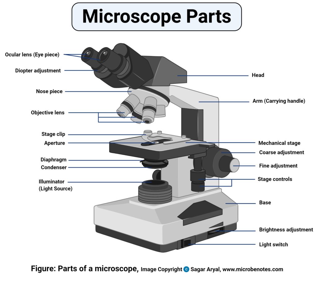
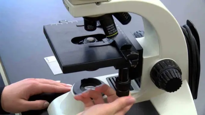
A microscope is an instrument used in viewing objects that are naturally considered too small to be seen with the bare eyes.
The science of investigating small objects using the microscope is referred to as microscopy.
Table of contents
History of the microscope
Although objects which look like lenses have evolve about 4000 years ago and there are Greek accounts of the optical properties of water filled spheres followed by many centuries documentaries on optics, the earliest known use of simple microscopes is traced back to the use of lenses in eye glasses in the thirteenth century.
The earliest discovered examples of compound microscopes, which combine the objective lens near the specimen with an eyepiece to view the real image appeared in Europe around 1620.
The name of the inventor is not known although there have been several claims over the years of its real inventor. Many of those claims are centered around the spectacle making centers in the Netherlands including claims that it was invented in 1590 by Zacharias Janssen.
Galileo Galilei, often referred to as the founder of the compound microscope seems to have discovered after 1610 that he could close focus his telescope to view small objects and after seeing a compound microscope built by Drebbel exhibition in Rome in 1624, built his own improved version.
Giovanni Faber formed the name microscope for the compound microscope Galileo Galilei handed over to the academia dei lincei in 1625.
Rise of modern light microscope
The first detailed account of the microscopic anatomy of organic tissue based on the use of the microscope was not discovered until 1644 in Giambattista odierna’s Locchio Della musca.
The microscope was still widely regarded as a novelty until 1660s and 1670s when it was used to study biology by naturalists in England, Italy, and Netherlands.
Marcello Malpighi an Italian scientist called the father of histology by some biological sciences historians began his first biological structures analysis with the lungs. Robert Hooke’s publication on micrographia in 1665 had a significant impact because of its illustration.
A meaningful input also came up from Antoine Van Leeuwenhoek who achieved 300 magnification using a simple single lens microscope. He sandwiched a very small glass ball lens between the holes in two metal plates riveted together and with an adjustable by screws needles attached to the mouth of the specimen.
Then Antoine Van Leeuwenhoek, discovered red blood cells and spermatozoa and also help spread the use of the microscope.
Types of microscope

There are generally several types of microscope namely.
- Compound microscope: This is the most popular and less expensive type of microscope used in the laboratories to carry out findings. They use lenses of different capacities to magnify small objects. This type of microscope usually consists of an eyepiece, a set of mirrors and the objective lens that function together. Here images are usually seen in two ways. With the help of the microscope camera the user can save the viewed images to make analysis or findings later on.
- Scanning Electron microscope: This type of microscope is able to detect objects with the 3-D imaging system. Here images have high magnification and resolution, but they appear in black and white. A specimen is coated in gold particles to produce a detailed image. This type of microscope is very important in viewing very tiny particles and images could be saved to be viewed later. Detailed images of bacteria, Viruses and other cellular components can be viewed by this device.
- Transmission Electron microscope: It uses electron to pass through a sample. In this microscope two varying images are produced by creating small slices with high magnification and resolution. This type of microscope is important in collecting images through the thickness. Of the specimen not just the surface of the object or specimen. These types of microscopes are found in the higher institutions and are very expressive.
- Stereoscope: objects in three dimensions are usually viewed by the stereoscope. It allows you to see details which can normally not be seen by the naked eyes even though they are not highly powerful like the other varieties of microscope. They are equally used in the dissection of small objects and so they are sometimes referred to as dissection microscopes.
- Fluorescence microscopes: Generally, microscopes are used to view varying image of a sample depending on how the image is formed. However, this type of microscope is used use specific colors of light to interact with dyes. It is used to view specific proteins within a cell. There is always a camera attached to it to, to capture images from the microscope.
- X-ray microscope: This type of microscope uses the electromagnetic radiation usually in the soft x-ray band to capture objects. It is relevant in tomography to produce three dimensional images of objects. Including biological materials that has not been chemically fixed yet
Uses of microscope in forensic science
Forensic science aids to know the past whether in terms of studying why a particular disease is spreading, carrying out an investigation on a past site of massacre. It’s also relevant in the legal profession in solving crime related matters.
In these aforementioned field the microscope is useful in reconstructing events of the past. Its uses in forensic sciences include.
- General criminal science: Evidences may make or break case when it comes to proffering solutions to a case. However, microscopes are important here because they can be used in investigative purposes and because they can magnify an object or a specimen to a very great extent. Microscopes can be useful in comparing the hair, fibers and other things recovered from the crime scene.
- Forensic epidemiology: The scientific study of the spread of disease is referred to as epidemiology. Forensic epidemiology usually has the same duty as epidemiology but in its case, it used for legal purposes. Forensic epidemiologists may be assigned to investigate dangerous bacteria like Salmonella. To carry out this task effectively, they make use of the microscope to study food for contamination. With the aid of the microscope, the presence of certain strains of bacteria will point out source of the contamination. This will literally help prevent more people from being affected and also point out the exact cause of the disease outbreak.
- Forensic anthropology: Here microscope is used to study tissues, bones or other things to investigate the cause of a death. Scanning electron microscopes are very relevant here, as they can be used to investigate the long-liquefied remains of a dead person in the soil.
- Forensic pathology: Forensic pathologists determine the way and manner in which person died through thorough investigations. If the person’s death was caused by a particular bacteria, forensic pathologists will now use the aid of a microscope to find out the nature and type of the bacteria. A microscope is also useful in determining the nature of the injury, to find out it was caused by a knife, bullet or something else.
More Reading…
References;
- Bardel David (2004)” The invention of the microscope.”
- The history of the telescope by Henry c king publisher courier Dover pp
- Murphy Douglas B Davidson Michael W (2011). Fundamentals of light microscope and electronic imaging (2nd ed). Oxford Willie Blackwell. ISBN 978-471-69214-0


