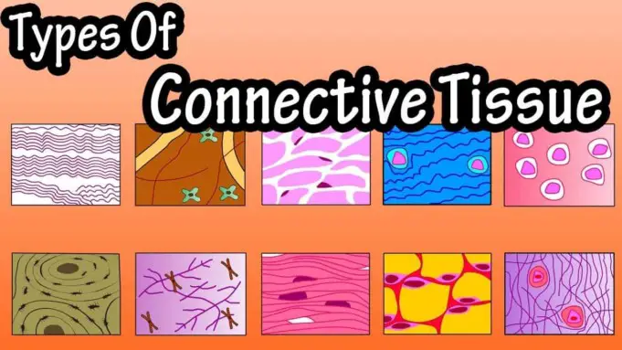
The term connective tissue was coined in 1830 by Johannes Peter Muller in German, Bindegewebe. The tissue was already recognized as a distinct class in the 18th century.
Connective tissue is one of the four significant tissue classifications (others are epithelial tissue, muscle tissue, and nervous tissue).
Connective tissue is found almost everywhere in the body, forming between other tissues and organs, between spaces, serving as a binding structure and providing metabolic support and protection. Its amount varies between organs.
Connective tissues are unique tissues that serve as frameworks, fill spaces, store fat, produce blood cells, protect against infections, and help repair damaged tissues.
Structure of Connective Tissues
Compared to other tissue types such as epithelial tissue, Connective tissue cells are farther apart, and the structure of the cells comprises an abundance of extracellular matrix between them.
This extracellular matrix is made up of protein fibers and a ground substance. The ground substance is a colorless, clear fluid consisting of non-fibrous protein and other molecules that fill the space between the cells and fibers.
The non-fibrous protein (proteoglycans and cell adhesion proteins) allows the connective tissue to act as the adhesive for the cells to attach to the matrix.
The ground substance of the extracellular matrix functions as a molecular sieve for substances to move between the blood capillaries and walls of cells.
The extracellular matrix consistency varies from fluid to semi-solid to solid, modified to suit various organs and conditions.
The extracellular matrix protein fiber provides support, and it is secreted by cells known as fibroblasts.
Fibroblasts produce three different types of connective tissue fibers:
- Collagenous fibers
- Elastic fibers
- Reticular fibers
Collagenous fibers
They are thick threads of the protein collagen. They are grouped as long, parallel bundles and are flexible but only slightly elastic. More importantly, they have significant tensile strength- they resist a considerable pulling force.
Thus, collagenous fibers are essential components of the body parts that hold structures together, such as ligaments (connectors bone to bone) and tendons (which connect muscle to bone).
Tissues containing abundant collagenous fibers are referred to as dense connective tissues. It appears white, and for this reason, collagenous fibers are sometimes called white fibers.
Elastic Fibers
They are composed of a protein called elastin. These are thin fibers that branch, forming complex networks. Elastic fibers are far weaker than collagenous fibers, but they stretch easily and resume their original length and shape after being tensed.
Elastic fibers are standard in body parts that are frequently under tension, such as the vocal cords, skin, lungs, arteries, and veins. They are sometimes called yellowfiber because tissues well supplied with them appear yellowish.
Reticular fibers
They are thin, short collagenous fibers with numerous branches that form delicate supporting networks in various tissue types, including the spleen.
You should know that when the skin is exposed to prolong and intense sunlight, connective tissue fibers may lose their elasticity, and the skin stiffens and becomes leathery. In time the skin may become sagged and wrinkled. Collagen injections may temporarily smooth out wrinkles.
However, collagen applied as a cream to the skin does not combat wrinkles because collagen molecules are far too large to get into the skin.
Type of Cells found in Connective Tissues
Connective tissue contains a variety of cell types. Some cells are called fixed cellsbecause they reside in the tissue for an extended time.
These include mast cells and fibroblasts. Other cells, such as macrophages, are referred to as wandering cells because they temporarily move through and appear in tissues, usually in response to an infection or injury.
- Fibroblasts are the most common type of fixed cell found in connective tissue. These large, star-shaped cells produce fibers by secreting proteins into the extracellular matrix of connective tissues.
- Mast cells are large and widely distributed in connective tissues. They are usually near blood vessels. Mast cells release heparin, which prevents blood clotting, and histamine, promoting some of the reactions associated with inflammation and allergies.
- Macrophage cell or histiocytes originates as white blood cells, and they are almost as numerous as fibroblasts in some connective tissues. Macrophages are referred to as phagocytes because they are specialized in carrying out a metabolic process called phagocytosis; that is a cellular process by which a cell uses its plasma membrane to engulf a larger particle, giving rise to an internal compartment called the phagosome. Macrophages can move about and function as scavengers and defensive cells that clear foreign particles from tissues.
Classification of Connective Tissue
Connective tissue is classified into two categories. First is the Connective tissue proper, which includes loose connective tissue and dense connective tissue.
Secondly are the specialized connective tissues, which are the cartilage, blood, and bone.
Loose Connective Tissue
Types of loose connective tissue include areolar tissue, adipose tissue, and reticular connective tissue. Areolar tissue forms delicate, thin membranes throughout the body.
These connective tissue cells are mainly fibroblasts, located some distance apart and separated by a gel-like extracellular matrix containing many collagenous and some elastic fibers secreted by fibroblasts.
Areolar tissue attaches the skin to the underlying organs, and they also fill spaces between muscles. It lies beneath almost all the epithelium layers, where its numerous blood vessels nourish nearby epithelial cells.
Adipose tissue or fatty tissues are formed when specific cells (known as adipocytes) store fat as droplets in their cell compartments (cytoplasm) and enlarge in size.
When adipocytes become so abundant, they tend to crowd other cell types and form fatty tissue.
Adipose tissue lies beneath the skin, in spaces between muscles, around specific joints. Adipose tissue cushions joints and some organs, such as kidneys. It also insulates beneath the skin, and it stores energy in fat molecules.
Reticular connective tissue is composed of thin, collagenous fibers in a three-dimensional network. It helps to provide the framework of specific internal organs, such as the liver and spleen.
Dense Connective Tissue
Dense connective tissue is a tissue that consists of many thick, closely packed, collagenous fibers in a fine network of elastic fibers. Dense connective tissues have few cells, most of which are fibroblasts.
Collagenous fibers of dense connective tissue are firm, enabling the tissue to withstand pulling forces. As parts of tendons and ligaments, dense connective tissue binds muscle to bone and bone to bone.
This type of tissue is also in the protective white layer of the eyeball and in the deeper skin layers. The blood vessels supplying blood to dense connective tissue are poorly established, thereby slowing tissue repair.
Cartilage
Cartilage is a rigid connective tissue. It provides support, frameworks, and attachments, protects underlying tissues, and forms structural models for many developing bones.
Cartilage extracellular matrix is abundant and is largely composed of collagenous fibers embedded in a gel-like ground substance. Cartilage cells, or chondrocytes, occupy small chambers called lacunae and lie completely within the extracellular matrix.
The cartilaginous structure is enclosed in a covering of connective tissue called the perichondrium. The perichondrium contains blood vessels that provide cartilage cells with nutrients by diffusion.
There is a lack of a direct blood supply to cartilage tissues, that is why torn cartilage heals slowly, and the cells do not divide frequently.
Different types of extracellular matrix distinguish three types of cartilage, they include:
- Hyaline cartilage: This is the most common type. It has very fine collagenous fibers in its extracellular matrix and looks somewhat like white glass. Hyaline cartilage is the type of cartilage found on the ends of bones in many joints, in the soft part of the nose, some part of the ribs, larynx, and in the supporting rings of the respiratory passages (trachea). Hyaline cartilage is also important in the development and growth of most bones.
- Elastic cartilage: On the other hand, has a dense network of elastic fibers and thus is more flexible than hyaline cartilage. It provides the framework found in the external ears and parts of the larynx.
- Fibrocartilage: This is a very tough tissue, has many collagenous fibers. It serves as a shock absorber for structures that are always subjected to pressure. For example, fibrocartilage forms a pad- intervertebral disc, between the individual bones of the spinal column. It also cushions bones in the knees and in the pelvic girdle.
Between ages thirty and seventy, a nose may lengthen and widens by as much as half an inch, and the ears may lengthen by a quarter inch because the cartilage in these areas continues to grow as we age.
Bones
Bone is the most rigid connective tissue. Its hardness is largely due to the high deposition of mineral salts, such as calcium phosphate and calcium carbonate, between cells.
The extracellular matrix also has many collagenous fibers, which are flexible and reinforce the mineral components of bones.
Bones internally support body structures, protect vital parts in the cranial and thoracic cavities, and are an attachment for muscles.
Bone also contains red marrow, which forms blood cells, and it stores and releases inorganic chemicals such as calcium and phosphorus. Bone matrix is deposited in a thin layer called lamellae, which form concentric patterns around tiny longitudinal tubes called central canals.
Bone cells are also known as osteocytes and are located in lacunae, which are rather evenly spaced within the lamellae. Consequently, osteocytes also form concentric circles.
In a bone, the osteocytes and layers of the extracellular matrix, which are concentrically clustered around a central canal, forms a cylinder-shaped unit referred to as osteon.
Many osteons are cemented together to form the substance of bone. Each of the central canals contains a blood vessel, which places every bone cell near a nutrient supply.
Also, bone cells have many cytoplasmic processes that extend outward and pass through very small tubes in the extracellular matrix called canaliculi. These cellular processes connect with the membranes and nearby cells.
As a result, materials can move rapidly between blood vessels and bone cells. Thus, in spite of its inert appearance, bone is a very active tissue that heals much more rapidly than cartilage.
Blood
Blood is a tissue that transports varieties of materials between interior body cells and those that exchange substances with the external environment, such as CO2 gas.
In this way, blood helps maintain stable internal environment conditions. Blood is composed of formed elements suspended in a fluid extracellular matrix called plasma.
The formed elements include red blood cells RBC (erythrocytes), white blood cells WBC (leukocytes), and cell fragments called platelets.
Most blood cells form in red marrow within the hollow parts of certain long bones, such as the humerus, femur, and ribs.
The function of Connective tissue
Below is a summary of the functions of the different type of connective tissue:
| Types | Function | Location |
| Loose connective tissue Areolar tissue Adipose tissue Reticular tissue | Bind organs and it holds tissue fluids. Protect organs, insulate the body, and store fat. Support | Beneath the skin, between muscles, beneath epithelial tissues. Beneath the skin, around kidneys, behind eyeballs, on the surface of the heart. Walls of liver and spleen |
| Dense connective tissue | Bind organs. | Tendons, ligaments, deeper layers of skin. |
| Cartilage Hyaline cartilage Elastic cartilage Fibrocartilage | Support protect and provide a framework. Support protects and provides a flexible framework. Supports protect and absorb shock. | Nose ends of bones ring in the walls of respiratory passages. The framework of the external ear and parts of the larynx. Between bony parts of the spinal column, parts of the pelvic girdle, and knee. |
| Bone Blood | Support protect and provide a framework. Transport substances and help maintain a stable internal environment. | Bones of skeleton Throughout the body, inside a closed network of blood vessels and in the heart chambers. |
Complications of connective tissue
A complication of connective tissue refers to disorders involving connective tissue that supports organs and other parts of the body.
The complications are numerous; however, they can be classified as:
Inherited Disorders of Connective Tissue
These are disorders as a result of gene mutation. Many under this class are quite rare. The following are some of the common ones:
- Ehlers-Danlos Syndrome (EDS)
- Epidermolysis bullosa (EB)
- Marfan Syndrome
- Osteogenesis Imperfecta
Autoimmune Disease
For other types of connective tissue complications, the cause is usually unknown. In some cases, researchers believe the disorder may be triggered environmentally for people who may be genetically susceptible.
In these complications, the body’s immune system produces antibodies that target the body’s own tissues.
These conditions include:
- Rheumatoid Arthritis (RA)
- Polymyositis and dermatomyositis
- Scleroderma
- Sjögren’s Syndrome
- Systemic lupus erythematosus
- Vasculitis
- Mixed connective tissue disease
Apart from the above mentioned, other complications of connective tissues that have been discovered include Scurvy, Fibromuscular dysplasia, myxomatous degeneration, and connective tissue neoplasms, including sarcomas such as hemangiopericytoma and malignant peripheral nerve sheath tumor in nervous tissue.
Sources
- Connective tissue; https://en.m.wikipedia.org/wiki/Connective_tissue
- Classification of Connective Tissue; https://www.histology.leeds.ac.uk/tissue_types/connective/connective_tissue_types.php
- Connective tissue; http://www.histologyguide.com/slidebox/03-connective-tissue.html
- Characteristics of Connective Tissue; https://courses.lumenlearning.com/boundless-ap/chapter/connective-tissue/


