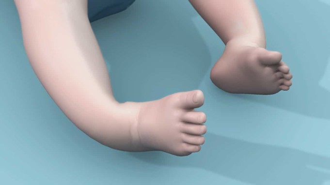
Club foot is a foot abnormality in which a child’s foot or feet are twisted inward or outward at birth. About average number of children with club foot have it on both legs.
This condition happens at birth hence it is also termed Congenital Talipes Equinovarus. A normally developing foot becomes a clubfoot during the second trimester of pregnancy.
A leg with club foot is observed to be twisted out of shape and position and if left untreated can result to difficulty in walking and pains.
Club foot is caused by an improper alignment of muscles to the bones of the leg. This is as a result of the tissues connecting the muscles to the bone (tendons) being shorter than usual.
This alignment involves the soft and bony structures in the hindfoot, midfoot and forefoot. Club foot can be mild or severe.
It can be treated successfully without surgery but in some severe cases, surgery is required. Club foot is the most common birth defect affecting the legs as it occurs in 1 to 4 of every 1,000 live births especially in firstborn children and males.
Cause of Club foot
The causes of club foot is unknown. It may be due to;
- Genetic factors
- Skeletal abnormalities
- Environmental factor
Genetic Factor
On genetic factor, changes in the genetic makeup (genes) might result to clubfoot. It may be hereditary as some parents have a history of having suffered such a condition in their childhood.
This increases the likelihood of giving birth to a child with the condition or deformity.
Skeletal Abnormalities
Another factor that may result in club foot is skeletal abnormalities such as spinal bifida; a condition whereby a developing baby’s spinal cord fails to develop properly as the spine and membranes around the spinal cord fails to close completely.
This causes a poor ability to walk. Hip dysplasia is another skeletal abnormality that can result in club foot. In this condition, the hip socket doesn’t fully cover the ball portion of the upper thigh bone leaving the hip joint partially or completely dislocated.
Both conditions occur mostly at birth. Club foot might also be due to environmental factor
Environmental factor
Factors like maternal age and mothers engaging in habits like smoking of cigarette and drinking of alcoholic drinks during pregnant may result to club foot or clubfeet in babies.
Although the actual cause of club foot is unidentified, the mechanism behind the condition is about the disruption of the muscle of the lower leg which leads to the shortening of the muscle or joint.
Symptoms of Club foot
The symptoms may include;
- Stiffness of the deformed foot
- Rotation of the foot or feet both inward/outward and downwards
- Difficulties in walking or disabilities
- Pain may be experienced in the later stage of life other than in childhood
- Wobbling while walking
- Affected leg may be slightly shorter than the unaffected leg.
Diagnosis of Clubfoot
A professional health care provider usually notice a clubfoot in a baby at birth. It can also be detected before birth via ultrasound exam in the 20th week of pregnancy.
The latter is easily detected if the feet are affected but no correction can be done until the birth of the baby. Regardless of how this defect is detected, more test is recommended to check for other health problems especially those related to the defect like spinal bifida and muscular dystrophy/ hip dysplasia.
A club foot is usually diagnosed through a physical examination whereby a thorough check is carried out on the baby on every part of its body at birth. An X-ray can also be carried out to detect the severity of the clubfoot.
Treatment
The treatment of club foot is carried out after the first or second week of the child’s birth. This is because of the flexibility and fragility of the baby’s bones, tendons and joints.
The treatment is of two approach; conservative and surgical approaches. Regardless of the approach used, the treatment is aimed at correcting the deformity thereby preventing long-term disabilities rendering it fully functional and pain-free.
Club foot can be treated in a combination of methods which includes;
- Stretching and casting also known as Ponseti method
- French method which involves realignment, taping, long-term home exercise and night splinting
- Surgery, which is not always necessary.
Ponseti method
The Ponseti method is developed by Dr. Ponseti, an Orthopedic Surgeon based at the University of Iowa in 1963 following an anatomical study of the foot.
Ponseti method is the most common treatment for clubfoot and it involves casting, Achilles tendon release and bracing.
In this method of treatment, the baby’s foot is manipulated into an improved or correct position and placed in a cast from the toes to the thigh to hold in position for about a week.
This procedure is repeated every week for about four to eight weeks. This re-position the foot by aligning the forefoot with the hindfoot. The casting procedure helps in correcting the cavus, adductus and varus deformities but the equinus which is corrected by Achilles tendon release (also called Achilles tenotomy).
In Achilles tendon release, the Achilles tendon is cut and after dressing the cut the leg is placed in a cast to fully correct the position. This is the longest cast lasting about three weeks.
The Achilles tendon regrow in a lengthened position during this time. Once a child’s foot is corrected or realigned, stretching exercise is advised for the care givers of the baby. The baby should be put in special shoes and braces also known as Foot Abduction Brace (FAB) as long as needed.
This may take up to three years. This helps prevent the foot from returning to the deformed shape. The brace is made up of two shoes or boot connected together by a bar. All these should be done according to the doctor’s directions for effective result.
Reoccurrence of club foot deformity is seen in some cases and this may be due to inadequate adherence to bracing. In other to correct this, the process of casting is repeated followed by surgery.
The chances of achieving an effective result is low compared to a casting done a week or two after child birth.
French Method
The French method of treatment is a physical therapy. The therapy is carried out daily for the first two months and thrice weekly for the next four months.
The foot is manipulated, stretched and taped using a non-elastic adhesive strapping to immobilize the foot. The muscle acting on the foot is stimulated by strengthening it to maintain the reduction achieved through manipulation before taping.
The child is to be engaged in home exercise on conclusion of the physical therapy and also splinting at night to maintain long-term correction.
Ponseti method is preferred to the French method as the latter prove less effective.
Surgery
Surgery is carried out when nonsurgical means of treatment fails to correct the deformity. The extent of the surgery depends on the severity of the deformity.
The surgery is carried out by an orthopedic surgeon on the child usually up to 9 to 12 months of age to correct all the component of the deformity (cavus, varus, adductus and equinus) at the time of surgery.
The surgeon can lengthen or reposition tendons and ligaments to help ease the foot into a better position. After surgery, the child is placed in a cast for about two months then a brace for a year or more to prevent recurrence of the deformity.
Prevention
There is no definite prevention for clubfoot as the cause is unknown. However, one can minimize the possible risk of giving birth to a child with clubfoot by abstaining from smoking and drinking of alcohol during pregnancy.
Also avoidance of the intake of drugs not prescribed or recommended by a doctor may be helpful in preventing clubfoot.
Epidemiology
Clubfoot occurs in 1 to 4 of every 1,000 live births especially in firstborn children and males. It is the most common birth defect affecting the legs. It most commonly affect people in low and middle-income countries (LMICs) with 80% or approximately 100,000 cases per year as of 2018.


