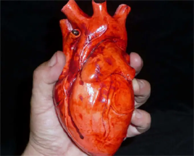
Normally, the heart is divided into two parts – left and right – which are separated by a septum (which separates the two upper chambers of the human heart).
The right side of the heart receives oxygen-free blood and directs it to the lungs. Blood saturated with oxygen returns from the lungs and enters the left side of the heart, and from there it goes to all other organs. The septum prevents the mixing of blood.
Some babies are born with a hole in the cardiac septum (on the upper or lower wall). The hole separating the upper chambers of the heart is known as an Atrial Septal Defect (ASD), and the opening in the lower part is a Ventricular Septal Defect (VSD).
In both cases (ASD and VSD), it turns out that the blood (enriched with oxygen and without it) is mixed. A large opening, in case of ASD, can cause an overflow of the lungs with blood, thereby complicating the functioning of the heart.
Before we proceed to understand this misfunction, let’s have a look at what is ASD and VSD:
Atrial Septal Defect (ASD):
Atrial septal defect (ASD) is an opening in the atrial septum, leading to shunting from left to right and overloading with the volume of the right atrium and right ventricle.
Children with ASD are rarely symptomatic but may experience long-term complications after 20 years which includes pulmonary hypertension, heart failure, and arrhythmias.
Adults and, less commonly, adolescents can suffer from intolerance to physical exertion, shortness of breath, fatigue, and arrhythmias due to this defect. Under ASD, soft meso systolic murmur at the upper-left edge of the sternum with a sharp and constantly bifurcated 2nd cardiac sound (S 2 ) is common.
The diagnosis is done by echocardiography. The treatment of this defect consists of using a transcatheter device for closure or surgery.
Ventricular Septal Defect (VSD):
Ventricular Septal Defect (VSD) is an opening in the interventricular septum, leading to communication between the ventricles. Large defects lead to a significant discharge of blood from left to right and cause shortness of breath during feeding and low growth rates during infancy.
Loud sharp holosystolic noise at the lower-left edge of the sternum is common among patients with VSD. Recurrent respiratory infections and heart failure may develop as a result of this defect.
The diagnosis is done by echocardiography. These defects may close spontaneously in infancy or require surgical intervention.
Facts:
- According to a report, about 5200 babies are born in a year with the hole in their hearts.
- Cardiologists classify ASD as a small cardiac abnormality.
- In some cases, when there are no severe symptoms that affect the quality of life, this syndrome can be perceived as an individual feature of the heart structure.
Causes of a hole in the heart
ASD and VSD are congenital heart defects. Typical causes of the hole are considered:
- Genetics: a child is at an increased risk of developing an Atrial Septal Defect if one of the parents has a congenital heart disease
- The presence of other genetic disorders: children with a hereditary disorder (for example, Down syndrome) often have congenital heart disease
- Smoking: children born to mothers who smoked during pregnancy are susceptible to various congenital heart defects.
Signs and Symptoms of a hole in the heart
Most children do not have any symptoms of ASD. Nevertheless, symptoms can occur at a more mature age – at 30 years or even later. Signs of ASD include:
- Heart murmur
- Fatigue
- Shortness of breath, palpitations
- Bluish skin color
- Swelling of the feet, legs, or abdomen
- Stroke
Symptoms of VSD (congenital heart defects) occur shortly after the birth of the baby – during the first few days, weeks or months. Among the symptoms are:
- Cyanosis, or a bluish tint of the skin, lips, and fingertips
- Fast breathing
- Poor appetite
- Swelling of the feet, legs, or abdomen
- Heart murmur (in some babies it may be the only sign of a defect)
Examinations necessary to confirm or rule out a violation
ASD and VSD are diagnosed in the following way:
- Physical examination (the doctor listens to the heart and lungs with a stethoscope to detect heart murmur)
- Echocardiography
- Electrocardiogram (ECG)
- Chest x-ray
- Cardiac catheterization
- Pulse oximetry
First, the doctor collects general data about the patient’s health, medical history, and other complaints. This will help to identify the causes and possible complications.
A physical examination is also carried out, that is, the doctor examines the skin, determines body weight, measures blood pressure, listens to heart tones.
Then a general analysis of blood, urine, biochemical blood analysis is prescribed. These studies help identify related diseases, cholesterol, and other important factors.
All this allows the doctor to accurately assess the health status of the patient, his/her heart, determine the size of the anomaly, and so on.
Available Treatment Options
In most cases, ASDs close on their own during the first year of the birth of the child. Based on regular examinations, a doctor may suggest treatment for a medium or large opening between the ages of two and five years.
Treatment usually consists of surgical procedures or catheterization, which allows you to “seal” the hole:
- Catheterization (performed under anesthesia) involves the introduction of a catheter into a vein in the groin and passage up to the septum. Two small discs that are attached to the catheter are pushed out – and close the hole between the atria of the heart. Over time, healthy tissue builds up around the device (over six months)
- The surgical operation allows the surgeon to close the hole with a special patch
A ventricular septal defect (VSD) is subject to simple control if it does not show any symptoms. In situations that require treatment, attention is paid to:
- Nutrition – special feeding or nutrition for children who are poorly developed. You may need breast milk, special supplements, a feeding tube or bottle for feeding.
- Surgery – Large hole in the heart requires open-heart surgery, which eliminates the opening in the septum.
Complications and Prevention
Of course, the likelihood and form of complications depend on many factors. But it’s important to understand that complications rarely occur. In fact, such diseases can develop:
- Renal infarction;
- Stroke;
- Myocardial infarction;
- Transient cerebrovascular accident
Safety Precautions
In order to maintain health, children and adolescents should be regularly examined by a physician to monitor the healing process after treatment with an ASCI or APS.
Both parents and children (who are undergoing the treatment) should strictly follow the doctor’s recommendations in order to gradually return to their usual daily life.
Precautions undergoing medical therapy are also of high importance in order to avoid some complications, infections such as catheter migration. This kind of outcome may occur during insertion when the catheter enters a side vein or reverse direction or from spontaneous migration at any time. That’s why hospitals, doctors, and nurses use professional neonatal Picc catheters, special vascular devices, etc.
The purpose of the article is educational and informational. The publication is not a substitute for personal specialist advice. If you have any health issues, consult your doctor now.
Author Bio:
Mahima is a content development specialist working with Credihealth. Her article is a combination of her journey of a healthy lifestyle and information from trusted websites. When she isn’t writing she can usually be found reading a good book.


