Abnormalities such as breast lumps are often detected by physical exams, ultrasound, mammography, and other imaging studies. Although it may not be possible to tell from these imaging tests whether the growth is cancerous or benign.
An ultrasound-guided breast biopsy, uses sound waves to locate abnormalities such as lump and remove a tissue sample from a suspicious area in the breast to be examined under a microscope.
Ultrasound-guided breast biopsy does not involve exposure to ionizing radiation like x-ray does. It is less invasive and leaves little or no scarring. It also helps to guide a radiologist’s instruments to the area of an abnormal growth in the breast.
Common Uses of Ultrasound-Guided Breast-Biopsy
An ultrasound-guided breast biopsy can be recommended by a doctor when a breast ultrasound that was carried shows abnormalities such as;
- Distortion in the structure of the breast tissue.
- A solid mass is suspected.
- An abnormal change in the breast tissue.
Ultrasound can serve as a guide to biopsy procedures such as;
- Core Needle: Also called automatic spring-loaded needle, is used to remove one sample of the breast tissue at a time or per insertion. It makes use of a large hollow needle.
- Wire Localization: This is used to assist or help a surgeon locate a lesion for a surgical biopsy. In wire localization, a guidewire is placed into the suspected area.
- Fine Needle Aspiration: It makes use of a very small needle which extracts cells or fluid from the abnormal or suspected area.
- Vacuum-Assisted Device: It is used to remove multiple samples of the breast tissue per insertion. It makes use of a vacuum-powered instrument.
Equipments used for the Procedure
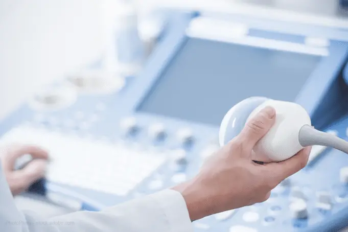
The types of equipment used for an ultrasound-guided involves a computer console, video display screen, and a transducer which sends and receives inaudible high-frequency sound waves that become visible on the visual display screen which looks like a computer monitor.
It also involves the use of one of these four types of equipment or instruments;
- Core needle or automatic spring-loaded needle
- Vacuum-assisted device(VAD)
- Guide wire (used for surgical biopsy)
- Fine needle aspiration
How you should prepare for the Procedure
When going for an ultrasound appointment in a hospital, you may not need any special preparations. However, here are a few tips to help you with your preparations;
- Always wear comfortable clothes and remove your jewelry.
- Ensure to give the doctor full details of your medical history before the needle biopsy.
- Adhere to all instructions given to you by the doctor before the biopsy. Some of these instructions may include; refraining from taking blood thinners, aspirin or some herbal supplements a few days before the biopsy.
- You should have a friend or relative accompany you especially if you have been sedated.
How Ultrasound and Ultrasound-Guided Breast Biopsy Works
Ultrasound scanners makes use of a computer console, a video display screen and a transducer. The transducer is a small device that is hand-held, it looks like a microphone and is attached to the video display screen. It is the transducer that send and receive sound waves that is made visible on the visual display screen.
A special gel is applied on your skin or a lubricant is applied on the transducer to prevent friction and enable high frequency sound waves to travel from the transducer through the gel into your body.
When an ultrasound probe is used to visualize the location of an abnormal tissue change, distortion, or mass, the radiologist inserts a biopsy needle through the skin to the targeted area and removes the tissue sample.
In surgical biopsy, an ultrasound probe is used directly guide a wire into a targeted area helping the surgeon to locate the area for excision.
Procedures of an Ultrasound-Guided Breast Biopsy
Ultrasound-guided breast biopsy is a minimal invasive procedure that is done on an outpatient basis. The procedure generally take one hour. Steps include;
- Lying down face up or slightly turned to the side on a table in the ultrasound room which is usually dark, to enable easy reading of the ultrasound images on the ultrasound screen.
- A local anesthetic will be injected into your skin, and more deeply into the breast.
- The radiologist or sonographer will press the transducer against the breast to locate the lesion.
- Once the lesion is located, a very small nick will be made in the skin at the point where the biopsy needle is to be inserted.
- The tissue samples will be removed using one of these three methods;
- A fine needle aspiration(FNA): which uses a fine needle and a syringe to withdraw clausters of cells or fluid from the breast.
- A vacuum-assisted device(VAD): which uses a vacuum pressure to pull tissue through a needle into the sampling chamber. The needle rotates its position and collects about eight to ten tissues from around the lesion area for sampling without reinserting the needle.
- Automatic spring-loaded needle: Is activated by moving the needle and filling the needle trough with cores of breast tissue. This process is done repeatedly for about three to six times.
- After the tissue sampling, the needle is removed.
- You may be asked to wait for a short while. Then the diagnostic radiologist will check if the imaging was complete. He will also check to see if both the imaging tests and the pathology explains one another.
- After an ultrasound-guided biopsy, patient are usually asked to go home.
For a surgical biopsy, its steps include;
- Lying down face up on a table in the ultrasound room which is usually dark, to enable easy reading of the ultrasound images on the ultrasound screen.
- A local anesthetic will be injected deeply into the breast to numb it.
- The radiologist or sonographer will press the transducer against the breast to locate the lesion
- When the lesion is found, the surgeon inserts a wire into the suspected area. At the biopsy site, a small marker may be placed to help locate the biopsy site in the future.
- Once the biopsy is completed, pressure is applied to stop any bleeding and the opening in the skin will be covered with a dressing.
- A mammogram may be used to confirm if the marker is in the right position.
Who Performs and Interprets the Results
A radiologist performs the ultrasound scan while a pathologist (a doctor who specializes in the examination of tissues and cells) examines the removed specimen(samples of tissue and cell) to make a final diagnosis.
The radiologist also evaluates the results of the biopsy to ensure that the image findings and the pathology explains one another.
Sometimes follow-up exams may be recommended to further evaluate any potential abnormality, to see if an abnormality has changed or it is stable, or to see if the treatment is working.
What You May Experience During and After the Procedure
There are some things you may experience during and after an ultrasound-guided biopsy which are completely normal.
During an ultrasound-guided breast biopsy
During a breast biopsy, you will be awake and must remain still while the procedure is going on.
You will feel little discomfort during the procedure. You will feel a pin prick and a stinging sensation when receiving the local anesthetic to numb the skin.
You may also feel pressure when the biopsy needle is inserted to collect sample tissues. It is normal to hear buzzing or click sounds from the sampling instruments.
After an ultrasound-guided breast biopsy
You may experience swelling which is usually temporary. Your doctor may recommend over-the-counter(OTC) pain reliever and ice pack for you.
Avoid any strenuous activity for at least a day. And ensure you contact your doctor if you experience bleeding, excessive swelling, redness, drainage or heat in the breast.
Benefits of an Ultrasound-Guided Breast Biopsy
Some known benefits of ultrasound-guided breast biopsy include;
- It does not use radiation.
- It is used to evaluate lumps near the chest wall or under the arm which may be hard to see with stereotactic biopsy.
- It is a less invasive procedure which leaves little or no scarring.
- It provides tissues and cells for sampling which can show whether a breast has lump or malignant.
- It is less expensive compared to other biopsy methods such as stereotactic biopsy or open surgical biopsy.
- Recovery time is brief or short and patients can resume their usual activities soon.
Risks/Complications of Ultrasound-guided Breast Biopsy
Unlike ultrasound which has no known risks, ultrasound-guided breast biopsy may have some risks because it involves penetrating the skin.
- Patients may occasionally feel significant discomfort.
- Bleeding and forming of hematoma. However the risk occurs in less than one percent of patients.
- Patient may be at risk of infection because the skin was penetrated.
- Sometimes the procedure may not provide the final answer to explain the abnormality.
Limitations of Ultrasound-Guided Breast Biopsy
A few limitations of this procedure is listed below;
- It may underestimate the extent of a present disease.
- Ultrasound-guided biopsy cannot be used if the lesion is not seen on an ultrasound scan.
- Surgical biopsy is usually recommended if the diagnosis remains uncertain.
- Clustered calcifications are nor clearly shown with ultrasound.
- Ultrasound-guided biopsy may occasionally miss a lesion or find it difficult to accurately target a very small lesion.


My sister would like to have a biopsy because there’s a lump on her left breast. It’s interesting to learn that a wire localization will be used to locate the lesion. I’d keep in mind to remind my sister that she would wear comfortable clothes before the ultrasound.