

There is a rare type of abnormal in-growth of cyst that can occur in anyone at the bottom of the vertebra column, very close to the tail bone (coccyx).
This growth is called a pilonidal cyst or sinus and the condition it results in the pilonidal sinus disease. Pilonidal is a skin infection which usually occurs as cyst.
The disease can present itself solo or with a couple of different other conditions and may be asymptomatic, but the most common form of pilonidal disease is seen in patients suffering from Erectile Dysfunction (ED); it is observed in this form as a painful, swollen lesion without an eye (opening) in the sacrococcygeal region about 4cm posterior to the buttock (anal orifice).
In some cases, spontaneous or unnoticed drainage of the cyst may occur in patient without their consent, prior to presentation to the doctor. Patients with pilonidal disease may present intermittent swelling and drainage of purulent, mucoid or even bloody fluid from the site; especially if the site has an opening.
The cyst usually contains hair, dirt and skin debris. It results I severe pain and can often become infected, it might ooze pus and blood and have a foul odor if punctured, in cases of no opening.
History Overview of Pilonidal Disease
Pilonidal Sinus Disease like most other disease condition has an uncertain history; disease conditions date back as far as the history of humans but if the event or occurrence of the disease is not recorded there is no way to ascertain its history.
Therefore, the beginning dates of many diseases as old as man is the date that they were written for the first time. For pilonidal disease, its date is 1833; Mayo Herbert, a British Physiologist, Anatomist and Surgeon (1796 – 1852), first described the disease as a ‘hair-filled sinus’ located in the sacrococcygeal region of women.
Later, an article was discovered, titled “Hair Extracted from an Ulcer” in the Boston Medical Surgical Journal which was published by Anderson in 1847. In the article, he reported the case of a 21-year-old young male with a scrophuloderma on his back.
In his article, Anderson reported to have drained the cavity after 3 weeks and a structure looking like a mesh made of multiple hairs of 2 inches long in the back of the patient and after complete drainage and cleaning of the hair in the cavity, the wound healed quickly. In 1854, seven years later, 3 other similar cases were reported by Warren.
This first series of reported cases and study formed the history of pilonidal sinus disease.
The disease was addressed with so many names such as sacral, coccygeal or sacrococcygeal infundibulum, dermoid and dermoid fistula, sacrococcygeal ectodermal sinus and congenital dermal sinus.
It was until 1880, that Hodges named the disease with a statement of “I venture to give the name of pilo-nidal sinus to this rather singular lesion.” He invented the name ‘pilonidal’ by conjoining the word “pilus” (Latin word: hair) and “nidus” (Latin word: nest).
Causes of Pilonidal Disease
Over 200 years ago, since the disease was first mentioned, there have been many fevered arguments and many theories to describe the etiology and whether the disease is congenital or acquired.
About 80 years ago, Gage reported that pilonidal cyst and sinuses are congenital, and he was supported. According to the congenital disease theory, it might have originated from caudal remnants of the neural tube, dermal inclusions produced by sequestrated epithelial structures or dermal tractions that are produced during the involution of the tail during embryonic development.
Pilonidal condition mostly affects men and is common in young adults. It is often familiar with individuals who sit a lot like taxi or cab drivers.
The origin of the growth of pilonidal cyst in the body is still very much unclear; some clinicians think that the ‘ingrown’ hairs are the cause for many of the pilonidal cases. Pilonial in Latin means “nest of hair” and doctors most times find hair follicles inside the cyst.
During World War II, more than 80,000 soldiers got pilonidal cysts that sent them to the hospital. Clinicians then thought they were because of irritation from riding in bumpy Jeeps. For a while, the condition was called “Jeep disease”.
Another theory proposes the reason to be due to trauma and long-term activities that affect causes friction that affect that particular region of the body, like sitting.
This group claims that these activities can force the hair growing in the area to burrow back under the skin; the body considers this hair foreign and launches an immune response against it, similar to how the body reacts when dealing with a splinter.
This immune response forms the cyst around the hair follicles; sometimes a person may have multiple sinuses that connect under the skin.
Also, an individual is likely to be affected with pilonidal disease if he/she were born with a small dimple in the skin between the buttocks. This dimple tends to get infected, though the reason for infection is not known but this is one of the known risk factors for individuals.
Risk Factors for Pilonidal Disease
Apart from those born with dimples in the buttock region, other risks factors that can make a person more susceptible to developing pilonidal cysts include:
- Obesity
- Large amount of body hairs
- Occupation requiring Prolonged sitting.
- Excessive sweating
- Male sex
- Stiff or coarse hair
- Inactive lifestyle
- Younger age (pilonidal cysts are most common in people in their 20s)
Signs and Symptoms of Pilonidal Disease
Pilonidal disease condition is mostly recognized when the region is infected and infection process may occur asymptomatically (not presenting any symptoms) or at first present a small, dimple-like depression on the surface of the skin.
However, once the depression becomes infected, it quickly evolves into a cyst; an enclosed sac filled with fluid, or an abscess; an inflamed tissue where pus gathers.
Symptoms of an infection may include:
- Swelling of the cyst just above the anus or near the tail bone
- Hair protrusions from the lesion
- Pain when sitting of standing.
- Redness, sore skin around the area
- Pus containing traces of blood draining from thee abscess resulting in a foul odor.
- Formation of more than one sinus tract/holes in the skin
- Low grade fever; but this is much less common.
Diagnosis of Pilonidal
Pilonidal cyst may be diagnosed by physical examinations by the doctor and by historical questioning about the condition, questions may entail:
- When did you first feel symptoms?
- Have you had a fever?
- Has the situation occurred before?
- What medications or supplements do you take?
Treatment of Pilonidal Disease
In conservative treatment, cases of pilonidal disease (cases which are asymptomatic) it is likely that the physician will prescribe a broad-spectrum antibiotic. Broad spectrum antibiotics are group of antibiotics that treat a wide range of bacteria.
Now it is important to know that this treatment does not heal the sinus tract, but it gives the patient a relief from the infection and discomfort.
Regular hair removal or shaving of the site, particular attention to hygiene and follow-up examination may be recommended by clinicians.
- Lancing is another treatment approach for pilonidal abscess or a collection of pus inside the sinus. Before this treatment process, local anesthetic is administered to the patient and then the doctor uses a scalpel to open the abscess. The doctor cleans away any hair, blood and pus from inside the abscess. The wound is sterilely dressed and allow to heal from the inside out, which takes up to four weeks and further treatment may not be required.
- Phenol injection is a type of treatment that involves the use of phenol (a chemical compound used as an antiseptic). In the course of this treatment, local anesthetic is administered, and phenol is then injected into the cyst. This procedure may be repeated several times. Eventually, in the course of this treatment, the lesion hardens and closes.
- This treatment has a high recurrence rate, and this makes it unfamiliar in most regions of the world. Doctors turn to surgery as treatment in most severe cases.
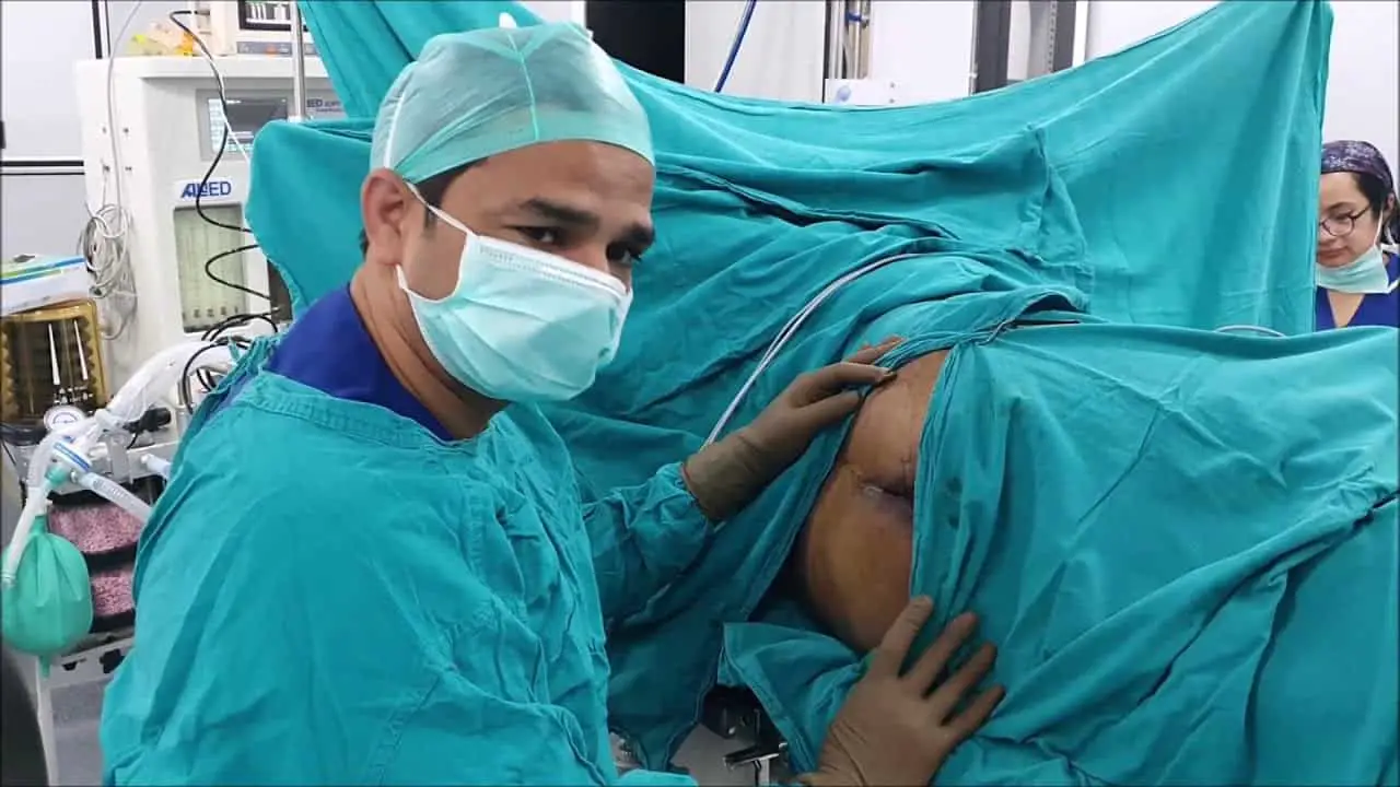
- Surgery is usually a course of treatment in cases of recurring pilonidal disease or if the case involves more than one sinus tract. Local anesthetic is also first administered during surgery, and then the lesion is opened by the surgeon, he then removes all the pus and debris. Once the procedure is completed, the surgeon will stitch the wounds closed. Dressings of the wounds after surgery may be needed to be changed and shaving of the site is recommended to prevent hair from growing into the wound.
Complications with Pilonidal Disease
There are some conditions termed complications that may arise for Pilonidal disease. These include wound infection and a recurrence of pilonidal disease even after surgery.
Some signs that the wound is re-infected include:
- Severe pain
- Inflamed and swollen skin
- Foul odor from wound
- Temperature of above 100.4oF
- Pus and blood seeping from wound site
If a chronically infected Pilonidal cyst is not treated or is not properly treated, the individual may be at slight risk of developing a type of skin cancer called squamous cell carcinoma.
Prevention and Management of Pilonidal Disease
Regular shaving of the area or use of hair removal products is highly recommended to reduce the risk of recurrence. To help prevent pilonidal cysts, individuals should try to:
- Avoid prolonged sitting.
- Regular exercising
- Keep the area clean on a daily basis.
- Avoid obesity.
Summary
Pilonidal disease is rare and not life threatening, the condition can be defeated with prompt attention and treatment to emerged symptoms and regular cleanliness and exercise to prevent the condition.
Sources;
- Pilonidal disease; http://fascrs.org/patients/diseases-and-conditions/a-z/pilonidal-disease
- Pilonidal disease; http://en.m.wikipedia.org/wiki/Pilonidal_disease
- Pilonidal Cyst and Sinus Clinical Presentation; http://emedicine.medscape.com/article/788127-clinical#b1
- Pilonidal sinus diseasehttp://www.ncbi.nlm.nih.gov/pmc/articles/PMC4607805/


")


