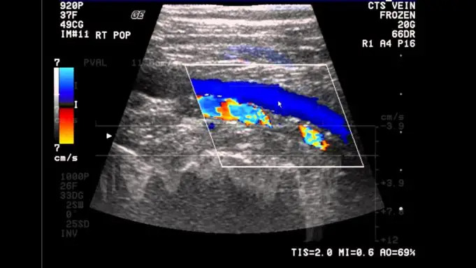
Venous ultrasound is a painless imaging test that is used to examine and produce real-time images of the veins in the body.
Venous ultrasound is also called Extremities ultrasound and is commonly used to diagnose conditions such as deep vein thrombosis (blood clot in the veins of the leg).
Ultrasound uses sound waves not radiation and can help to detect vein abnormalities and guide placement of a needle into the vein.
Uses of Venous Ultrasound
Since ultrasound scan are used to produce real-time images of the internal organs/structure, Venous ultrasound can be used to;
- Identify abnormal blood flow and damaged valves.
- Determine the cause of swollen legs due to long-standing.
- Guide placement of catheter or needle in the vein.
- Bypass a blocked blood vessel and map out veins in arms or legs that may be removed during a surgery.
- Examine if a blood vessel grafting used for dialysis is functioning as expected or not.
In infants it helps to diagnose conditions such as;
- Blockages such as blood clot.
- Increased blood flow which may be caused by an infection.
- Reduce blood flow to some organs.
- Evaluate and determine the connection between a vein and an artery which is seen in arteriovenous malfunctions.
Who Performs the Venous Ultrasound
Venous ultrasound is performed by an ultrasound technologist or technician. A diagnostic radiologist or radiologist is a medical professional who is trained to interpret ultrasound images.
They specialize in interpreting medical imaging tests including MRI scans, ultrasound and CT scans.
How does ultrasound works?
Ultrasound scanners make use of a computer console, a video display screen and a transducer.
The transducer is a small device that is hand-held, it looks like a microphone and is attached to the video display screen. It is the transducer that send and receive sound waves that are made visible on the visual display screen.
A special gel is applied on your skin or a lubricant is applied on the transducer to prevent friction and enable high frequency sound waves to travel from the transducer through the gel into your body.
Procedures of Venous Ultrasound
Venous ultrasound is performed in a hospital. The procedure of the Venous ultrasound takes less than 30-45 mins generally. Steps include;
- Lying down face up on a table in the ultrasound room which is usually dark, to enable easy reading of the ultrasound images on the ultrasound screen.
- Depending on the type of examination, you may be told not to eat or drink anything except water for about 6-8 hours before the ultrasound.
- A special water-based gel will be rubbed on the area to be examined to reduce friction and help the transducer slide across your skin. It is the transducer that sends and receives sound waves to produce the image on the ultrasound screen.
- The technologist will occasionally press the transducer down to enable the technologist to have a clear imaging that will help diagnose your condition.
- The water-based gel is then wiped off from your skin.
- You may be asked to wait for a short while. Then the diagnostic radiologist will check if the imaging was complete.
- After an imaging test, the patients are usually asked to go home.
What you may experience during and after a Venous ultrasound
During a Venous ultrasound, you may experience minimal or no discomfort as the transducer presses against your skin.
After the Venous ultrasound, you will be asked to dress and sit for a while as the result is being reviewed. You may be given the results or asked to return to the hospital in a few days and should be able to resume normal activities after an ultrasound scan.
Benefits of Venous Ultrasound
Some known benefits of Venous ultrasound are listed below:
- It is not painful although it may be uncomfortable temporarily.
- It is noninvasive meaning there are no injections or needles.
- It helps to detect and show blood movements within blood vessels.
- It is less expensive and easy to use compared to other imaging methods.
- It is widely available compared to other imaging methods and provides great internal details when accessing soft tissue structures.
- It can be used to detect blood clot in the veins especially of legs(from the thigh down to the knee) before they dislodge and pass to the lungs.
- It is completely safe and it uses sound waves not radiation.
- It provides a more clear picture of soft tissues that did not show up clearly on x-ray.
Risks/complications of Venous Ultrasound
Although standard ultrasound diagnostics do have a known harmful effect on the human body, but an interpretation of the ultrasound may lead to other procedures such as biopsy, aspiration, and followups.
Limitation of Venous Ultrasound
Though ultrasound scan is painless and has no known harmful effect on the human body, venous ultrasound may find it difficult to see veins that lay deep beneath the skin especially calf veins which are very small.
Conclusion
If a venous ultrasound is recommended for you by a doctor, you need not to get worried or scared over it because it is a noninvasive medical exam that does not require needles. It is completely safe and painless.


