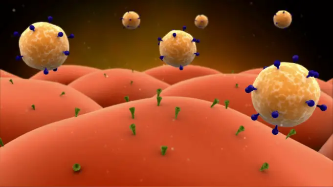
Membrane proteins exist in the cell membrane and allow for the communication between the cell and the extracellular space. The use of recently developed purification methods has revealed just how complex their structure is and has brought to light the crucial role that membrane proteins play in the different chemical pathways that build our metabolic architecture.
Receptors and Messengers
Singer and Nicolson first postulated the fluid mosaic model of cell membrane in the early 1970s (1). In their model, they established that lipids, proteins, and carbohydrates were the main components of the cell membrane and that proteins played a crucial role in the transmission of information from the outside to the inside of the cell and vice versa (1). In fact, these amphiphilic molecules can be external or embedded in the lipid bilayer matrix where they are known as intrinsic or integral.
Intrinsic proteins are generally membrane molecules that behave as receptors. These elements, known as first messengers, mediate signal transduction to extracellular stimuli, which triggers a cell response (2).
Part of the receptor is exposed to the external space of the cell and has a binding site for the ligand. Ligands include hormones, lipoproteins, transferrins, neurotransmitters, and other molecules (2). Once the ligand reversibly binds to the external domain of the receptor, the intracellular portion of the transmembrane receptor activates proteins such as kinases, guanylate cyclase, ion transporters, G proteins.
This initiates a cell response to the initial stimulus (2). The stimulus is transduced to the interior of the cell through second messengers. Second messengers can be classified into three classes:
Hydrophilic/cytosolic: localized in the cytosol, including cAMP, cGMP, Ca2+ and involved in phosphorylation mediated responses. Their main targets are protein kinases (3).
Hydrophobic/membrane-associated: localized in intermembrane spaces and involved in regulation of kinases and phosphatases, transcriptional factors, and G protein associated factors. Some examples include DAG, PIP3, arachidonic acid and ceramide (3).
Gaseous: localized in the cytosol and within the cell membrane. Some examples include nitric oxide and carbon monoxide. They activate cGMP (3).
The cellular response in signal transduction cascades involves the synthesis or inhibition of targeted proteins mediated by the expression of effector genes. Response to stimulus will not only involve gene expression but also cell proliferation or apoptosis.
Importance of Effective Protein Purification Methods for Gene Therapy
Identification of many receptors is often not an easy task. This is mainly because they are present in very minute amounts within the body. However, modern accurate technology that analyzes protein purification has provided researchers with more detailed information about structure, affinity and stability of receptors. New equipment can determine structural integrity of receptors in different conditions.
This valuable input, in addition to the capacity of isolating genes encoding receptors for specific ligands, has allowed scientists to design and understand the complex chemical pathways in our bodies (4).
Several metabolic and endocrine diseases are associated with an impaired interaction between a chemical signal and its specific receptor. Therefore, understanding the chemical networks of receptors, including the first and second messengers involved, is allowing for better diagnoses and treatment therapies for many diseases (5).
That is the case with gene therapy, which has been showing continuous progress and has created positive outcomes for a wide range of genetic diseases such as hematological, ocular, and neurological disorders as well as cancer (6).
There is an urgent need for better scientific research to lead the way for the development of better phenotypic correction techniques with gene therapy so that we can replace palliative treatments.
SOURCES:
- Lombard J. Once upon a time the cell membranes: 175 years of cell boundary research. Biol Direct. 2014; 9: 32. Published online 2014 Dec 19. doi: 10.1186/s13062-014-0032-7
- Yeagle P. The Membranes of Cells (3rd Edition). Academic Press (2016) ISBN: 978-0-12-800047-2.
- Kenneth B. Storey (1990). Functional Metabolism. Wiley-IEEE. ISBN 0-471-41090-X
- Lodish H, et al. Molecular Cell Biology. Section 20.2: Identification and Purification of Cell-Surface Receptors. New York: W. H. Freeman; 2000.
- Gerok W. Cell receptor defects as the cause of endocrine and metabolic diseases (author’s transl). Klin Wochenschr. 1979 Jun 15;57(12):613-23
- Kumar S, et al .Clinical development of gene therapy: results and lessons from recent successes. Mol Ther Methods Clin Dev. 2016; 3: 16034. Published online 2016 May 25. doi: 10.1038/mtm.2016.34.


