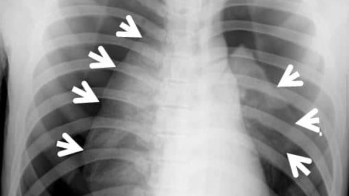
Pneumothorax is an abnormal medical condition characterized by the accumulation of air in the pleural cavity or pleural space.
The accumulation of air (pneumothorax), reduces the lungs capacity to expand this, causing difficulty with breathing and also the accumulation of air in the pleural cavity may result in the increase of tension in the space.
Pleural itself, is a bi-layer mucous membrane that enclose(surrounds) the lungs. It is further sub- divided into two parts which is the visceral part and the parietal part.
The visceral part is the party of pleural that surrounds the lungs (lining the chest wall) while the parietal part is the continuation of visceral part that surrounds the thoracic wall (lining the outer surface of the lungs).
The pleural cavity exists between the lungs and the chest wall or thoracic wall. So, the pleural cavity or pleural space can be said to be a space between the parietal pleural (lining the chest wall) and the visceral pleural (lining the outer surface of the lungs).
The accumulation of air(pneumothorax) reduces the lungs capacity to expand thus, causing ‘difficulty with breathing’. Also, the accumulation of air in the pleural cavity may result in the increase of tension in the space.
In normal adult, the pressure that exist in the pleural cavity (intra-pleural pressure) is always negative. This negativity is what keeps the lungs inflated and promote breathing.
However, the pressure becomes positive in pneumothorax which can cause atelectasis (collapse of the lungs) and this can lead to dyspnea (shortness of breath).
The intra-pleural pressure is generated by the fluid that is not ally present in the pleural cavity or pleural space. This helps to lubricate the visceral and parietal part of pleural.
Pneumothorax can also lead to a gradual or progressive oxygen shortage and low blood pressure. If the progressive oxygen shortage and low blood pressure is not reversed as soon as possible or almost immediately, it can be very fertile.
Types of pneumothorax
Pneumothorax is classified into two types;
- Spontaneous pneumothorax
- Non-Spontaneous (traumatic) pneumothorax
Spontaneous pneumothorax
This is called spontaneous pneumothorax because it does not occur after injury. It is further sub divided into two depending on whether there is an underlying problem in the lungs.
- Primary spontaneous pneumothorax: This occurs with no underlying lung disease and it can also be termed Idiopathic cause of pneumothorax (unknown cause). This type of spontaneous pneumothorax can occur in a healthy adult. Nevertheless, some risk factor may include: Smoking, family history, body type (slim or tall) etc.
- Secondary spontaneous pneumothorax: This occurs with an underlying lung disease. Anyways, older people are at a higher risk in this type of pneumothorax than the young people because of the underlying problem in the lungs. Some underlying lung disease include:
- Chronic obstruction pulmonary disease (COPD) which may account for approximately 70% of pneumothorax.
- Infection of the lungs which may occur in the case of tuberculosis.
- Cancer of the lungs
- Chronic obstruction airways disease seen in severe asthma and cystic fibrosis
- Connective tissue disease such as rheumatoid arthritis (RA).
- Interstitial lungs disease as seen in the case of idiopathic pulmonary fibrosis.
Non-Spontaneous(traumatic) pneumothorax
This exist after a trauma. Trauma like RTA (road traffic accident), Stab injury or may follow surgical procedures like lungs biopsy, central line placement, poorly primed chest tubes etc. Traumatic pneumothorax is sub divided;
- Open pneumothorax: This is characterized or developed after an injury in which an open communication is developed. The open communication is between the pleural cavity and outer part of the body. Air enters into the pleural cavity during inspiration (breathing in) and also comes out during expiration (breathing out) in the course of respiration (breathing in and out).
This can cause collapse if the lungs(atelectasis) , also may result in hypoxia ( reduction in concentration in oxygen), hypercapnia ( high concentration of carbon dioxide in the blood), dyspnea( shortness of breath) and asphyxia ( loss of consciousness due to interruption of breathing).
- Closed pneumothorax: This occurs during a mild injury. During a mild injury, air enters into the pleural cavity, then the hole in the pleural is sealed and closed. This condition does not produce hypoxia (low concentration of oxygen). The air from the pleural cavity is absorbed slowly.
- Tension pneumothorax: Though tension pneumothorax occurs in both spontaneous and traumatic pneumothorax. But it is more likely to occur with traumatic pneumothorax because it is a life-threatening condition.
This is an injury to the chest wall which causes hole in the chest. The hole sometimes is covered by a light or thin tissue which may behave like a flapping valve.
It is called a flapping valve because it allows the entrance of air into the pleural cavity or pleural space during breathing in but prevent the exit of air during breathing out due to its nature.
Due to this event (entering of air into the pleural cavity or pleural space and no exit of air during expiration), the pressure that exist in the pleural cavity (intra pleural pressure) increases normally.
This condition is very fertile as it can cause total collapse of the lungs.
Causes of pneumothorax
Pneumothorax (air in pleural cavity) may be as a result of damaged if injury to the thoracic wall, chest wall or lungs. This damages or injury can be during an accident, bullet or stab injury as mentioned in traumatic pneumothorax.
However, the types of pneumothorax listed above, are base in the causes. Hence the cause are as follows:
- Trauma: This is a serious injury that occur due to RTA (road traffic accident), chest injury as a result of stab, puncture, wounds, blunt chest trauma, gunshot wounds etc.
- Lung infection: Lungs infection can cause tuberculosis, pneumonia, lung abscess etc.
- Catamenial in relation to menses in girls (chest endometriosis).
- Iatrogenic: This is by following surgical procedures like lung biopsy, central line placement and also chest surgery (thoracotomy).
- Idiopathic cause of pneumothorax is unknown, but this is closely related to primary spontaneous pneumothorax.
- SCD (sickle cell disease) can also cause pneumothorax because the patient has something similar to primary spontaneous pneumothorax. This is because some air sac under the pleural (sub pleural blebs) can rupture. That is, burst open into pleural space and cause primary spontaneous.
Other causes may include;
- Measles
- Inhaling of foreign body
- Emphysema (destruction of elastic recoil of the lungs and loss of alveolar wall).
Symptoms of pneumothorax
Pneumothorax has some which will make the doctor to help detect what is really wrong. These includes:
- Difficulty with breathing and shortness of breath: This is the commonest symptom of pneumothorax.
- Chest pain: Most patient that goes to the hospital always makes a complain about the chest. After a while the patient might start feeling tightness of the chest or steady ache in the chest region.
- Cyanosis: This is a bluish discoloration if the skin and sclera due to low oxygen content of blood.
- Tension pneumothorax can be a symptom because if the low blood pressure, fast its rapid pulse rate. This may make the patient unconscious.
- Cough: This can also be a symptom of pneumothorax.
Treatments of pneumothorax
Treatment is either conservative where in:
You do a chest physiotherapy and incentive spirometry while giving oxygen support if necessary and treating the cause like in secondary spontaneous pneumothorax. This helps for the absorption of the excess air and resolution.
And is best when the accumulated air is small and the Pneumothorax is not in tension (this means that the air does not have an escape route, enters the pleural space but can’t leave).
- Surgery: For Tension Pneumothorax or massive Pneumothorax (plenty of air in the pleural space), surgical intervention is necessary.
- Needle aspiration: Under emergency, a needle is first inserted into the chest (2nd intercostal space, the space between second and third ribs, tracing it to the middle of the clavicle). This helps to drain out some air.
Then a Chest tube is inserted and connected to a bottle called underwater seal to help to remove the remaining air. This procedure is called Closed thoracotomy tube drainage.
Conclusion
Conclusively, pneumothorax can heal itself only of these is a small amount of air that is trapped in the pleural space or pleural cavity. And also, if there is no underlying lung disease concerning the patient.
Secondary spontaneous pneumothorax should be attended to immediately because of the underlying lung disease and Tension pneumothorax should also be taken seriously because it is a life-threatening condition.
If it is not treated with immediate effect, it can cause to atelectasis (collapse of the lungs) which can lead to death. Checkout our post on hemothorax.


