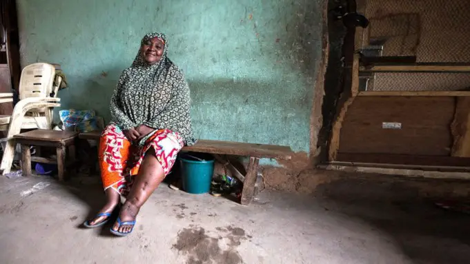
Lymphatic filariasis, commonly known as Elephantiasis, is a neglected tropical disease caused by the parasitic nematodes or roundworms of the family Filarioidea.
These thread-like worms are transmitted to humans from mosquitoes. There are sometimes no visible symptoms. However, it can be characterized by severe swelling in the arms, legs, breasts, or genitals.
This also causes the skin to become thicker and painful and also potentially limiting a person’s social and economic capabilities.
The type of worms that cause lymphatic filariasis are Wuchereria bancrofti, which is responsible for the majority of the cases; Brugia malayi, and Brugia timori are also responsible for the remainder of the cases.
These worms are capable of staying in the body for a long time and damaging the lymphatic system. Diagnosis is performed by testing the blood for antibodies against the disease.
Lymphatic filariasis can be prevented by treating the entire affected community in a mass deworming exercise. This is to be done annually for six years to get rid of the disease population. Antiparasitics can also be used to prevent the disease from spreading further.
According to the Centre for Disease Control and Prevention, the disease affects over 120 million people in 72 predominantly sub-tropical countries, which include Africa, Asia, the Western Pacific, and some regions of South America and the Caribbean.
The United States had its last known case of the disease in Charleston, South Carolina. Lymphatic Filariasis has long since disappeared from the region as of the early 20th century.
The infection is classified as a neglected tropical disease and is one of the four main worm infections of which includes Onchocerciasis, also known as river blindness.
History of Lymphatic Filariasis
The condition is thought to be as old as 4000 years. Possible symptoms of elephantiasis have been discovered from ancient Egyptian and West African artifacts.
The first description of lymphatic filariasis was in gree literature were scholars tried to differentiate between the symptoms of the condition and those of leprosy.
Jan Huyghen van Linschoten – a dutch merchant and historian – in the 16th century first documented the symptoms of the disease during the exploration of Goa.
Timothy Lewis, In 1866 made the connection between microfilariae and elephantiasis, establishing the course of research that would ultimately explain the disease. In 1876, Joseph Bancroft was the first to discover the adult form of the worm.
In 1877, Patrick Manson postulated that the lifecycle which involved a vector. He further proceeded to demonstrate the presence of the parasites in mosquitoes.
Although, he incorrectly hypothesized that the disease was transmitted through skin contact with water in which the mosquitoes had laid eggs.
There was no valid theory of transmission until 1900 when George Carmicheal Low determined the mode of transmission by discovering the presence of the worm in the proboscis of the mosquito vector.
Causes and Risk Factors of Lymphatic Filariasis
Wuchereria bancrofti, which is responsible for the majority of the cases of the disease, while Brugia malayi and Brugia timori cause most of the remainder of the cases.
Adult filarial worms nest in the lymph vessels, mate, and produce millions of microfilariae that circulate in the blood during their lifetime.
The average lifespan of the worms is about 6-8 years. Mosquitoes become infected when they bite and suck the blood of an infected host. These microfilariae grow into infective larvae in the mosquito.
The parasite is then transmitted to another person when the vector bites the skin when it passes into the bloodstream and into the lymphatic vessels where they mature into adult worms.
Several species of mosquitoes are responsible for transmitting the parasite, although this depends on the geographical location.
The common vector for the parasite is the Anopheles mosquito in Africa, and in the Americas, it is Culex quinquefasciatus. The Aedes and Mansonia can transmit the infection in Asia and the Pacific.
Several bites from mosquitoes over months to years are required to get the infection. People living in tropical or subtropical regions over a long period, especially in areas where the disease is common, are at the most considerable risk of getting infected.
Signs and Symptoms of Lymphatic Filariasis
Most of the cases of lymphatic filariasis are asymptomatic. This means they show no visible symptoms of the infection.
However, these infections can still cause significant damage to the lymph vessels and the kidney; they can also make changes to the body’s immune system.
The most apparent sign of lymphatic filariasis occurs when it develops into elephantiasis (thickening of the skin or tissues) limbs or scrotal sac, or lymphoedema (swelling of the tissue).
The breasts and genitals can also be involved. These deformities can lead to social stigma, low body image, and less than ideal mental health.
It can also cause one to be unable to participate in social and economic activities. Isolation and poverty are common for those with these body defects.
The body’s immune system’s response to the disease can also cause episodes of local inflammation on the skin and lymph nodes. This can result in secondary bacterial infection of the skin due to the impaired immune system and damage to the lymphatic system.
These acute episodes can be severe and may last for weeks, weakening the sufferer and reduce their ability to make a living.
Diagnosis
Diagnosis occurs by identifying the type of microfilaria in the blood smear using a microscopic examination. This may prove difficult as the microfilariae circulate only at night (this is also known as nocturnal periodicity).
Thus, blood should be collected at night when the parasite circulation is at its peak. The blood smear should be prepared and stained with Giemsa stain.
Examining the blood serum for antibodies against the disease may also be used as a method of diagnosis lymphatic filariasis.
Treatment
Treatment of the infection is dependent on the region of the world where the disease was acquired.
For example, albendazole is used along with ivermectin to treat the disease in sub-Saharan Africa, while albendazole is used with diethylcarbamazine citrate (DEC) in the rest of the world.
Antibiotics such as doxycycline are also useful in treating the infection.
It, however, takes a longer time (about 4 to 6 weeks) for effective treatment compared to anthelmintic drugs like albendazole. It is also not to be used in young children and pregnant women.
Prevention and Control of Lymphatic Filariasis
The World Health Organization recommends mass drug administration or mass deworming.
This would involve treating entire groups of people who are at risk with a single annual dose of two medicines, namely albendazole along with either diethylcarbamazine citrate or ivermectin.
The drug regimen would depend on the occurrence of other filarial diseases and would be administered at least twice a year.
Consistent treatment using all three drug combination would cause the worms to die out within weeks compared to the months or years it would take to clear out using only a combination of two drugs.
Other methods preventing the disease include using insecticide-treated nets and fumigating areas prone to mosquito infestation.


