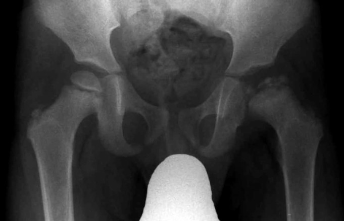
Legg-Calvé-Perthes Disease, or simply Perthes disease, is a rare disease condition that frequently affects children’s hip joints. The femoral head (the long bone of the thigh), which refers to the “ball of the hip,” is mostly affected.
The femur head is attached to the pelvic in the “ball and socket” hip joint; when a person develops Perthes disease, and this joint dies from lack of blood supply.
Perthes disease condition develops when there is a temporary loss of blood supplied to the hip joint (head of the femur), which causes the entire bone to die, known as avascular necrosis or Osteonecrosis (death of bone cells, osteocytes) occurs. A blood supply delivers necessary nutrients and oxygen to parts of the body, including bones.
A shortage in this regular supply may cause adverse effects to that location of the body and, as in Perthes disease, which is like starvation of the bone.
The weakened bone (femoral head) gradually breaks apart and may lose its round shape. The body system may eventually heal and restore blood supply to the ball, and the ball heals.
But if the femoral head is no longer preserves its original shape after it heals, it can cause pain and stiffness of the joint. The complete process from bone death to bone restoration can take several years.
Like most other medical discoveries, the name Legg-Calvé- Perthes disease was gotten from the three doctors who instrumentally unveiled the condition and its treatments.
In 1910, the Perthes disease was recognized and published as a condition unrelated to tuberculosis by three doctors working independently. Arthur Legg (1874-1939), Jacques Calvé (1875-1954), and Georg Perthes (1869-1927) were the three physicians associated with the discovery of the disease.
Although it has been originally agued to be first officially discovered by Dr. Georg Perthes, an Orthopedic Surgeon (branch of medicine that deals with the prevention, correction of disorders of the bone, and associated muscles and joints) from Germany, and X-ray diagnostic pioneer, in 1910.
The first debate suggests that Dr. Perthes took the first X-ray of a patient with this newly discovered hip condition. The second row of debaters could not place Dr. Perthes as the “Father of the Disease” firstly because, in 1909, Henning Waldenström (1877-1972) described the disease, but associated it with tuberculosis and secondly, because around that period of Dr. Perthes X-ray diagnostics, Dr. Jacques Calvé (1875-1954), a French Orthopedic Surgeon, and Dr. Arthur Legg (1874-1939), an American Orthopedic surgeon, also published findings of the disease.
While these doctors officially identified the condition that ultimately came to bear all three names, the disease was first described by an Austrian doctor named Karel Maydl in 1897.
But because of additional research and publications by other Orthopedics, particularly Dr. Perthes, the disease became medically known as the Legg-Calvé-Perthes Disease.
It is about a century in history to the discovery of the disease, and efforts in research and treatment of this idiopathic avascular necrosis of the capital femoral epiphysis of the femoral head is still an ongoing global one, as reflected in its international nature of the official name: Legg-Calvé-Perthes disease.
It took the effort of four genius-doctors from Germany, America, France, and Austria to first raise awareness about this disease.
Risk factor of Legg-Calvé-Perthes Disease
The risk factors for Perthes disease may include:
- Age. Perthes disease can affect children of any age, but it has been recorded most commonly to begin between ages 4 and 10.
- Sex. Perthes disease is four times more common in males than females.
- Race. White northern Europeans appear to be affected more frequently than all other races, though a lack of reliable epidemiology may exist in the Southern Hemisphere (Asians, Eskimos, and Caucasians).
- Genetic Mutations. For an insignificant number of persons, Perthes disease appears to be associated with mutations in certain genes, although more study is needed.
Epidemiology
Perthes disease is one of the common hip disorders in young children, occurring in an estimated figure of about 5.5 of 100,000 children per year, with both hips affected in about 15% of the population.
In a lifetime, the risk of a child developing the condition is about 1 per 1,200 individuals. The United Kingdom incidence rates show an interesting pattern with low incidence rates in London and a progressive increase of the disease in more northern regions, maximal in Scotland.
Some research indicates an increase in the prevalence of the disease in socioeconomically deprived communities of developed countries. One possible explanation that is considered to be the cause of this prevalence is exposure to tobacco smoke.
However, this is significantly confounded by the strong socioeconomic gradient common to both smoking and Perthes disease.
Causes of Legg-Calvé-Perthes Disease
Perthes disease is a hip disease that develops due to a complete shortage in or too little blood supply to the ball portion of the femur head (hip joint).
This portion of the femur is known to be the growing portion (epiphysis) of the bone’s upper end. Without enough blood supply, the bone-forming cells (osteoblasts) and resident bone cells (osteocytes) degenerate, and the bone becomes soft, weak, and tends to fracture easily.
The thigh bone (the upper end that cleaves to the pelvis) becomes fragile as bone mass is lost. This fragile area may fracture from the inside, causing a deformity. A thin line of the bone’s decreased density may be visible on the epiphysis, which may represent such a fracture within the bone.
The damaged bone may become fragmented and cause irregularities when blood flow to the affected area eventually resumes. As the bone re-grows and re-ossifies, it may become deformed, resulting permanently in the upper thigh bone’s malformation, for instance, unusually enlarged or abnormally flattened epiphysis.
The Perthes disease remains an idiopathic condition because the cause of the reduced or complete shortage of the blood supply to the hip joint is still unknown.
Some recent research indicates that there might be a genetic link to the disease, but more studies and research are needed to verify this claim.
Symptoms of Legg-Calvé-Perthes Disease
One of the major symptoms is Limping. As the ball of the thighbone flattens, it can make walking difficult. Other symptoms may be due to shortage of blood supply to the entire bone, they include:
- Pain or stiffness of the hip, groin, thigh, and knee.
- Decreased muscle strength of the thigh.
- Inflammation and irritation in the hip area may result in muscle spasms.
- Decreased range of motion of the hip joint.
- Pain that worsens with activity and improves during rest.
Perthes disease mostly affects one hip joint in children, but in some cases, usually at different times, it has bilaterally presented itself.
Diagnosis of Legg-Calvé-Perthes Disease
Diagnosis of a Child with Perthes disease begins with a physical examination (physiotherapy) to determine the range of motion within the hip joint. However, additional testing is very necessary to confirm Perthes disease.
These confirmation tests may include a bone scan, MRI, and X-ray. All three imaging tests are used to observe damages to the affected area’s bone and tissues.
When Perthes disease is diagnosed in a child, the doctor (orthopedic) will likely order periodic X-rays to monitor the disease’s progression. The images also help the physician determine the effectiveness of the treatment.
Other tests may also be conducted periodically to cross-check the disease’s progress. These include measuring the child’s thigh for indications of muscle atrophy (loss of muscle tissue).
Treatment of Legg-Calvé-Perthes Disease
Perthes disease treatment and management depend on the stage of the child or ward’s condition and the patient’s age. The treatment period often requires immobilization, methods to keep the child’s hip joint from moving, and limits daily activities.
For younger children, an orthopedic specialist (a doctor specializing in deals with the prevention, correction of disorders of the bone and associated muscles and joints) may recommend a wait-to-see approach, which involves treating with possible medications weight-bearing restrictions. Older children may be either treated surgically or non-surgically.
These non-surgical approaches of treatment in older children may include the use of anti-inflammatory medications, such as ibuprofen (Avil, Motrin), Naproxen sodium (Aleve), and physical therapy.
Some patients may need to walk with aids such as crutches, wheelchairs, or prescribed bed rest during the treatment process. In cases where Perthes disease progresses, the next step of action may be to place the child in a cast to keep the hips in the best healing position. Generally, patients younger than eight years old can be treated without surgery.
If the condition does not improve, a doctor may recommend a surgical approach. The older the ward, the more likely they will need surgery.
There are different types of surgery that can improve a Perthes disease condition. Some surgical procedures may involve removing the particles that restrict hip joint movements. While other surgeries conducted involves molding the entire portion of the femur.
The surgeon can also move the hip and femur to improve their alignment, a surgical procedure known as an osteotomy. After either surgical procedure, the child or ward is placed in a cast for several weeks to protect the alignment.
After removing the cast, physical therapy will be needed to regulate blood circulation, restore muscle strength, and restore motion range. Monitoring of the hips with an X-ray through this final stage of healing may continue.
Home care therapy is important alongside medical treatment. Activities involving light stretches can improve pain in the hip, and the child may also use heat pads or ice packs.
Over-the-counter prescriptions to relieve pain are also recommended by doctors during this healing period. However, it is advised to refrain from high-intensity activities, such as running and jumping, generally are not recommended due to the stress they place on the hip joint and thighs.
Complications of Legg-Calvé-Perthes Disease
The primary and seemingly only complication of Perthes disease in children is the increased risk of developing Arthritis later in their adult lives.
In general, young children with Perthes disease after the age of 6 or more are likely to develop hip problems later in their life. The younger wards have a better chance of complete healing with no future complication due to the bone’s longer re-ossification time.
As children below 6 have fewer ossified bones than those above 6, this allows for a longer healing gap of 2 years to reform the hip joint.
Summary
According to AAOS (American Academy of Orthopedic Surgeons), the general prognosis for most children with Perthes disease is very good. Within a two-year space of treatment, most wards recover and return to their daily activities.
Sources;
- Perthes Disease; https://orthokids.org/Condition/Legg-Calve-Perthes-Disease
- Perthes Disease; https://www.ouh.nhs.uk/paediatricorthopaedics/information/conditions/perthes-disease.aspx
- Perthes’ Disease; https://patient.info/bones-joint-muscles/hip-problems/perthes-disease
- Perthes Disease (Legg-Calve-Perthes Disease); http://www.hopkinsmedicine.org/health/conditions-and-diseases/perthes-diseaase-leggcalveperthes-disease


