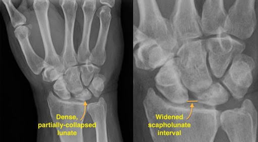
Kienbock disease is a disorder involving one of the major bones of the wrist called the lunate. The lunate bone is one of the eight carpal bones in the wrist; located at the middle of the base of the wrist that articulates (joins) with the radius of the forearm.
History of Kienbock Disease
In 1843, a close description of the Kienbock disease was first presented by Peste in the French medical literature, the description of the condition involved the collapse of the carpal lunate.
Some decades later, Robert Kienbock, a Viennese Radiologist in Austria (1871-1953), termed the condition “lunatomalacia”, a disease which later bears his name.
This Kienbock disease was observed by Dr. Robert disrupts the blood supply to the lunate by rupturing the ligaments and blood vessels around the lunate, which caused the bone to fracture with subsequent damage.
Later in 1928, another physician, Hulten, deducted from his examinations, an association between Kienbock disease and the presence of negative ulna variance (an abnormal formation of the ulna bone).
He continued progressively in the treatment by advocating for shortening of the radius. Thereafter, other researchers presented a contrary option of lengthening the ulna bone to restore the differential ulna variance.
Stages of Kienbocks Disease
In an early stage, kienbock’s disease may present severe pain. The disease rarely affects both wrist but as time passes and the condition progresses, the tissues of the bone may die leading to more pain, wrist immobility and arthritis.
A 2014 extensive study on the disease revealed that the disease’s progressive rate varies from case to case and from stage to stage.
Kienbock’s disease progresses through four stages:
Stage 1 of Kienbock Disease
- The initial stages describe abnormal or improper blood flow to the lunate carpal
- Damages may not be revealed by an X-ray scan of that area of the hand
- Sensation on the wrist become painful and it might be misinterpreted for a sprain
Stage 2 of Kienbocks Disease
- The bone is hardened due to total loss of blood supply
- This condition is called Sclerosis stage and it can be seen through an X-ray scan
- The wrist becomes swollen, tender and extremely painful to touch
Stage 3
- The lunate carpal dies and breaks apart, causing a shift of position of the wrist bones
- Pain sensations persist and motion is limited accompanied with a weakened grip
Stage 4
- The bones surrounding the lunate begins to deteriorate(worsen).
- This may lead to arthritis of the wrist
- Without immediate medical intervention, the disease can be debilitating (loss of strength or energy) in this stage.
Symptoms of Kienbock Disease.
Damages to the lunate can result to stiffness (wrist immobility) and severe pain. As it progresses, the injury can also lead to wrist arthritis.
Some common symptoms shown by patients of Kienbock’s disease are as follows:
- Swelling
- Wrist pain
- Tenderness of the skin area over the lunate carpal
- Stiffness of the wrist
- Weakened hand grip
- Difficulty in turning the hand
- Clicking sounds when the wrist moves.
Causes of Kienbock Diseases
There is no definite cause of Kienbock’s disease; at least a primary cause of the condition is still unknown. Speculations about a number of factors that can predispose a person to kienbock’s disease have been drawn but it is well known due to lack of evidence that the disease is hereditary, but it is possible that unidentified and verified genetic factors could contribute to the development of the condition.
Kienbock’s disease is also, often associated with injury to the wrist and hand such as trauma or accident that affects vessels supplying blood to the lunate.
Repetitive micro-injuries to the wrist such as a prolong use of a jackhammer, skeletal variations: the ulna bone may be shorter than the radius bone which may cause issues, also the lunate bone may be irregular and this place it at risk.
These conditions may be associated with:
People who are at Risk of Kienbock’s Disease?
- Kienbock’s disease can be found more commonly in people who have medical conditions that affect blood supply. It is associated with diseases like lupus, sickle cell anemia and cerebral palsy.
- People with difference in the length and shape of their forearm bones; the radius and the ulna. Putting more pressure on the lunate carpal.
- Patients with only one blood vessel supplying blood to the lunate instead of the usual two vessels. This affects blood supply to the bone.
- Kienbock’s disease occurs mostly in men between 20 and 40 years, especially those engaged in regular heavy manual labour.
When to visit a doctor?
If Kienbock’s disease is not treated, the lunate bone will continue to deteriorate; this may lead to severe pain and loss of the movement in the wrist.
In the early stage of the disease, conservative treatments may be able to relieve the pain. When the wrist pain persists over a reasonable period of time, it is important to have it checked out as early diagnosis and treatments can lead to a better outcome.
Diagnosis of Kienbock Disease
What kinds of test will the doctor use to ascertain your condition?
The physician attending to any Kienbock’s disease related case will ask for a medical history, lifestyle including occupation and other particular questions about the wrist pain. A physical examination will first be conducted on the wrist and hand and an X-ray scan may follow to further examine the wrist bones.
As earlier explained in the aforementioned stages of Kienbock’s disease, early Kienbock’s disease does not reflect on an X-ray. Therefore, an MRI (Magnetic Resonance Imaging) or a CAT (Computed Axial Tomography) scan can be carried out instead, to examine blood flow to the wrist.
Kienbock’s disease is a slow-progressing condition and patients most times have the condition for months before they seek treatment. This can make it difficult to diagnose in its earlier stages.
Doctors in the aspect of differential diagnosis, on imaging consider Ulnar Impaction Syndrome; a sclerosis change at the proximal ulnar aspect of the lunate.
Treatments for Kienbock Disease
The treatment of Kienbock’s disease totally depends on the stage of the disease when it is discovered. Medical operations to attempt restoration of blood supply to the lunate bone may be performed.
Conservative management with anti-inflammatory drugs, rest and immobilization in mild cases is very effective. Hand therapy does not change the course of the disease, but it can help minimize loss of motion from the disease.
The most common surgical therapy with exceptional positive results is radial shortening to correct negative ulna variance. Other medical procedures includes;
- Revascularization: This is a medical grafting procedure that entails grafting a piece of bone and blood vessels from another bone in the arm or hand to the lunate in order to restore blood flow. A piece of metal in the wrist (external fixator) may be used to keep the graft in place and relieve pressure on the lunate.
- Arthropasty: Total wrist joint replacement, the lunate is replaced with an artificial bone made either of silicon or pyrocarbon. Though this procedure is not frequently used, the method is also known as lunate excision.
- Metaphyseal core decompression: This medical procedure involves leveling the forearm bones by scraping the two bones involved without removing any bone tissue.
- Joint leveling: This is used to stop the disease from progressing when the two forearm bones are not the same length. The procedure involves removing a section of the longer bone (usually the radius) or grafting a piece of bone onto the shorter bone (usually the ulna). This removes the pressure on the lunate.
- Capitates-Shortening Osteotomy: Removal of a piece of another wrist bone, the capinate, and fusing it with other segments of the same bone. It is very effective at the early stages of kienbock’s disease, combined with revascularization.
- Inter-carpal fusion: The lunate is fused to adjoining bones to create a solid bone. This medical procedure relieves pain and allows partial wrist motion.
Any of the above procedures are also effective. In refractory cases (cases that are strongly opposing to conservative treatments), Proximal Row Carpectomy (PRC) is used as a salvage procedure.
Summary
Recovery time from surgery might be up to five months and the patient may be required to wear a cast to immobilize the wrist during healing. Also, physical therapy can be of great help in maximizing the use of the wrist with proper movement and strengthening exercises.
Sources
- Kienböck’s Disease; http://www.medicalnewstoday.com/articles/264720
- Kienbock’s Disease; http://www.assh.org/handcare/condition/kienbocks-disease
- Kienböck’s Disease; https://radiopaedia.org/articles/kienbock-disease-2#nav_treatment-and-prognosis
- Kienbock Disease; http://emedicine.medscape.com/article/1241882-overview#a1
- Kienbock’s Disease; http://www.physio-pedia.com/kienbock%27s_Disease


