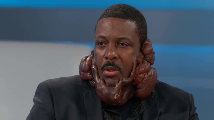
Keloids is a swelling. It looks like fibrous swelling around the skin. It looks as if it is a scar. The problem is only manifest in humans. It looks like a tumor and it manifests because of a dense fibrous tissue overgrowth in humans.
The problem is a form of the healing process and it manifests when the skin is recovering from the injury healing process. Though it is a healing, it is not a normal one. It is rather an excessive healing process of skin injury.
During the process of healing, human tissue grows above the borders of the healed wound. Furthermore, it could regress, but that is not spontaneous. The problem can recur after sometimes but that is after excision.
This is not the same thing with a hypertrophic scar. The difference between the two is that in the case of hypertrophic scar, erythematous is evidence. This simply means that it is not visible in dark-skinned individuals.
Furthermore, it is also pruritic. When means that it is fibrous lesions. This does not often grow beyond the initial injury. It does not grow that original boundary of that injury. It could be subjected to more partial spontaneous resolution.
Hypertrophic scars would always occur following a thermal injury. It can manifest as a result of other forms of injuries especially those injuries that have to do with the deep dermis. It can be said that the keloids are bumps.
What are the causes?
It is not easy to identify any etiology as the cause of that problem. Furthermore, this condition is not linked with any kind of gene. However, because of the peculiar way the problem manifest, experts are beginning to agree that it may have some genetic roots.
Research has so far linked the conditions to some genetic linkages like Blood Group A, HLA-DR5, HLA BW35, HLA-DQW3 as well as HLLBW16 and HLA-B21, HLA-B14 and so on.
There are some known causes of this condition. Skin trauma is often fingered as the cause of this problem. The trauma can manifest in different ways, which include physical and pathological.
When it is physical, it manifests in the form of surgery, and earlobe piercing. When it is pathological, it manifests in the form of chicken pox and acne and so on.
These are the major causes of keloids so far. However, the availability of foreign elements to the body as well as hematoma, infection and increased skin tension could cause that problem.
In some individuals particularly hypertrophic scar could be susceptic. There are at least two modes of inheritance which include the recessive and dominant inheritance are factors to the problem.
What are the clinical features of the condition?
Keloids are available all over the globe and the problem is more pronounced in women than it is men because of the ears piercing. Furthermore, the problem is more among African women particularly black women.
Among the dark pigments’ racial groups, this condition is more pronounced than in other groups. Research has shown that keloids are more manifest among the darker skinned individuals.
This means that it is more in black Africa. The second is the Asians and followed by the Caucasians. The condition is not normal in albinos and other races.
Furthermore, the problem is manifest in almost all the age groups but is more among the newly born and the elderly people. The highest incidence of this condition is among those individuals that are aged ten to thirty years.
Furthermore, research has shown that the condition can manifest in 5 to 15 percent of wounds. The areas of the body the problem often occurs include the abdomen, back, chest, breast, lower extremities as well as the neck, face, and the ear lobes.
Keloid is reported to have occurred on the sole, but that is not common. It can occur above the skin level and it goes beyond the wound margin.
When it manifests, it could continue to grow for a year, and it can shrink a bit. When it is growing, it can grow beyond the skin level. When it manifests, it is always painful, vascular and it can be itchy.
When it is beyond the growth stage, it maturates and resolute. It can be flat and broad. At this time, it has stopped growing. During pregnancy, it is common for the problem to become manifest as it can begin to grow again.
Complications
There are some complications that are associated with the problem and here are some of them.
- It comes with infection
- Ulceration from trauma is certain
- It leads to malignant change. Because of that, it could lead to fibrosarcoma condition.
- It can lead to itching and the tenderness
Treatment
So far there is no satisfactory treatment for this condition. This is because there is no single method of dealing with the problem.
Some of the measures adopted to deal with that problem include compression therapy, occlusive dressing as well as intralesional corticosteroid injections, cryosurgery, excision as well as radiotherapy and even laser therapy.
Excision method could account for fifty to one hundred percent. Surgery alone is not the best way out of the problem.
Another way to treat it is excision and radiotherapy. One of the treatment options includes the surgical excision and this is followed by the superficial irradiation. This can be done within 24 hours after surgery. This solution is not the final way out of the problem.
Excision and triamcinolone injection
excision should be followed by triamcinolone injection. The treatment option is followed for six months. Research has shown that this method is more successful than those already discussed.
Triple therapy
this is another treatment option and it also includes surgical excision. It is followed by radiotherapy and this takes place within 24 hours.
After that, another medication which triamcinolone injection is is applied. This method seems to be more successful when compared to such other methods like radiotherapy and surgical excision.
Compression therapy
this is another treatment option, and this involves the application of consistent controlled pressure to the affected part of the body especially the wounded area.
During the healing process, the mechanical force can be applied, and this interferes with that process and this often results in a flat scar.
There are other treatment options such as:
- Button therapy on ear lobes
- Zinc oxide adhesive plaster on facial keloids
- Application of ear clip
Furthermore, experts have recommended silicone cream or occlusive dressing. In addition to that, silicone gel could be applied to deal with that problem.
There are other innovative treatments such as bleomycin, intralesional interferon as well as doxorubicin and 5FU and verapamil and several others. Finally, the scar could be allowed and monitored as it regresses.


