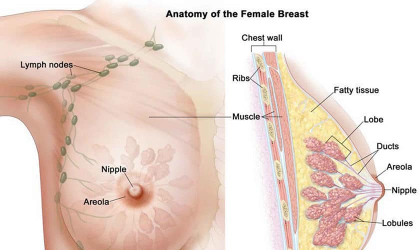
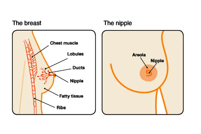
The intraductal papilloma is a breast condition where wart-like lump or tumour develops in one or more of the milk ducts in the breast. These lumps or tumour are made up of fibrous tissues, glands and blood vessels.
In the affected milk ducts, abnormal and atypical cells can be found. These cells are not cancer cells, but they increase the risk of developing breast cancer in the future. Some people who have multiple intraductal papillomae may also have a slightly higher risk of developing breast cancer.
The intraductal papilloma occurs close to the nipple but also likely to happen anywhere else in the breast area. The intraductal papilloma is a benign (a breast condition) not cancer. However, many people tend to confuse it with papillary breast cancer because they have similar names.
The Intraductal Papilloma mostly occurs in women between the age of 35 and 55. It also occurs in men but rarely. The intraductal papilloma develops as the breast ages and changes.
Types of intraductal papilloma
There are different types of intraductal papilloma, and they are:
- The Solitary Intraductal Papilloma: This occurs when a single tumour or lump grows in the milk ducts. It is felt as a lump close to the nipple. Nipple discharge or bleeding may manifest. This type of intraductal papilloma is not associated with a higher risk of cancer.
- Multiple Papilloma: Just like the name suggests, multiple papillomae occurs in clusters of little lumps. This type of intraductal papilloma has been associated with a slightly higher risk of cancer. This is because multiple papillomae have been linked to a precancerous breast condition called the Atypical Hyperplasia.
- Papillomatosis: This condition is grouped in with intraductal papilloma. It is associated with a higher risk of breast cancer. Papillomatosis occurs when there is an abnormal growth of cells in the milk ducts.
Symptoms
The intraductal papilloma is not usually painful, although some women can experience pain and discomfort in their breast. Other symptoms include;
- Small lump or multiple small lumps
- Breast enlargement
- Nipple discharge (without pressure applied to the breast)
- Bloodstained fluid from the nipple
Diagnosis

Generally, the intraductal papilloma can be found by chance during breast routine check-up. For an accurate diagnosis, the doctor may recommend the following test be carried out.
Mammograph
Mammographs is done by using low-energy x-rays to examine the breast. Mammographs also known as mastography, uses doses of ionising radiation to creates pictures. The images produced are analysed for abnormal characteristic masses.
Breast Ultrasound
Breast ultrasound involves the use of sound waves to produce images of the internal structures of the breast. This scan is used to diagnose lumps, tumours and other abnormalities in the breast.
Breast ultrasound is known to be more effective in detecting The Intraductal Papilloma than a mammograph. This is safe as it doesn’t use any radiation, and it is non-invasive.
Breast Biopsy
To rule out cancer, the doctor will carry out a breast biopsy. It involves inserting a thin needle into the breast tissue and extracting some cells. This type of breast biopsy is referred to as the fine-needle aspiration.
If there is discharge from the nipple, the doctor might perform a surgical biopsy. This will enable them to examine the breast tissue thoroughly to check for cancer cells.
Ductogram
A ductogram is a type of x-ray. A contrast dye is injected into the breast ducts so the doctor can view them in the x-rays. This is used to determine the underlying cause of nipple discharge.
Treatment of intraductal papilloma
The intraductal papilloma doesn’t dissolve on their own, but they can be removed through surgery. Procedures such as the following can be adopted to remove the intraductal papilloma.
Excision biopsy
This procedure involves the removal of the papilloma or the affected part of the milk duct. The surgery is usually conducted under local anaesthesia.
There may be a small wound from the incision and a scar, but the scar will fade over time. The tissues removed during the procedure are also further examined to confirm the diagnosis and check for cancer cells.
Vacuum-Assisted Excision Biopsy
This procedure involves sucking out the affected tissue – a hollow probe connected to the vacuum device through a small cut made on the skin.
With a mammograph used as a guide, the affected breast tissue will be sucked through the probe by the vacuum into the collecting container.
This is done under general anaesthesia. Some bruising may occur but will resolve in a few days. The tissues collected under this procedure are also further investigated. Further surgery may also be required if, after the above procedures, the patient continues to have nipple discharge.
The doctor may suggest Microdochectomy (removal of the affected duct or ducts) or total duct excision (removal of all major ducts). Breastfeeding will not be possible after a total duct excision but may still be possible after the removal of the affected ducts.
Although surgery should solve the challenge, if the nipple discharge starts again, another operation will be required. The patient may need to have more ducts removed.
After surgery, there may not be any reason to consult your health care provider. Although patients with multiple intraductal papillomae or whose intraductal papilloma contained abnormal cells might have followed up appointments with your doctor if you suspect anything due to certain noticeable changes in your breasts.
Prevention
There is no particular way to prevent intraductal papilloma. Although, you can increase the chances of early detection by being breast aware, having annual mammography and having a physical examination by a doctor.
The sooner the diagnosis is being made, the better your chances at beating it. You can also contact your doctor if there are any concerns about your breast health.
Sources
- Intraductal Papilloma; Healthline
- Intraductal papilloma: What you need to know; Medicalnewstoday
- Central Intraductal Papilloma; Breastcancer


