Cancer happens when cancerous cells in the body begin. Cells in almost any part of the body can become destructive and can spread to other regions.
Gallbladder cancer usually starts in the gallbladder, and to understand this type of cancer, it imperative to know about the gallbladder and its functions.
The gallbladder is a tiny, pear-shaped organ beneath the Liver: Where they are both located behind the right lower ribs. In an average human, the gallbladder is usually about 3 to 4 inches long and typically no more extensive than an inch.
The gallbladder collects and stores bile, which is a fluid manufactured in the liver. Bile aids digestion of fats in foods as they pass through the small intestine. Bile is produced by the liver, where it is transported into pipes that move it to the small intestine for use or stored in the gallbladder and released later for use.
When food (fatty food) is being digested, the gallbladder secretes and delivers bile via a small pipe called the cystic duct. The cystic duct is conjoined with the hepatic duct (which comes from the liver) and forms the bile duct.
The bile duct joins with the duct from the pancreas (the pancreatic duct) to discharge into the first part of the duodenum at the ampulla of Vater.
The gallbladder helps digest food but is not essential for living as a lot of people remove their gallbladder and go on to live healthy lives as its not life-threatening.
Types of gallbladder cancers
Gallbladder cancers not common, and most types of cancer are usually adenocarcinomas. Adenocarcinoma is cancer that starts in gland-like cells that line many exteriors of the body, including the surface of the digestive system.
- Papillary adenocarcinoma or papillary cancer is an unusual type of gallbladder adenocarcinoma whereby the cells in these gallbladder cancers are arranged in finger-like projections. Generally, papillary carcinomas are not likely to spread into the liver or closeby lymphatic nodes. They usually tend to have a better prognosis outlook than most other types of gallbladder adenocarcinomas.
- Other kinds of cancer can start developing in the gallbladder, such as squamous cell carcinomas, and carcinosarcomas, adenosquamous carcinomas, but are very rare.
Gallbladder cancer is not common. When gallbladder cancer is at its early stages, the odds of getting a cure is very high. But most gallbladder cancers are not discovered at an early stage when the prognosis is still good.
Gallbladder cancer is not easI[y diagnosed because it often shows no distinct indication and, the hidden nature of the gallbladder makes it difficult for gallbladder cancer to be diagnosed.
Symptoms
Gallbladder cancer signs and symptoms may comprise:
- Nausea or vomiting
- Itchy skin
- Abdominal bloating and Abdominal pain, especially in the upper right part of the abdomen
- Lumps in the stomach
- Dark urine
- Fever
- Jaundice
- Greasy or Light-colored stools
- Drastic weight loss usually as a result of the loss of appetite
Causes
A chronically swollen gallbladder is a prevalent link among a sizeable number of the risk factors for gallbladder cancer.
For instance, when a person has gallstones, the gallbladder may secrete bile more slowly, which means that cells in the gallbladder are unprotected from the chemicals in bile for a longer time. This could result in swelling and irritation.
In another instance, irregularity in the ducts that move fluids from the pancreas and gallbladder to the small intestine may allow fluid from the pancreas to reflux into the gallbladder and bile ducts.
This backward flow of pancreatic fluid may inflame and incite the development of the cells lining the gallbladder and bile ducts, which can heighten the chances of developing gallbladder cancer.
Experts and researchers are beginning to realize how risk factors like swelling may drive inevitable transformation in the DNA of cells, which allows them to grow out uncontrolled and become cancers.
DNA is the substance in each of our cells that makes up our genes, the guidance for how our cells perform. We resemble our mother and father because they are the origin of our DNA. But DNA affects more than our looks.
Genes regulate when cells grow and divide into new cells, and perish. These genes are known as oncogenes. Genes that slow down meiosis or result in cell mortality at the right time are referred to as tumor suppressor genes.
Cancer can be induced by DNA mutations that stimulate oncogenes or turn off tumor suppressor genes. Changes in many different genes are usually required for a cell to turn cancerous.
Some people get DNA mutations from their parents that vastly increase the risk for particular cancers. But hereditary gene mutations are not believed to induce many cases of gallbladder cancers.
Gene mutations linked to gallbladder cancers are often acquired during one’s lifetime rather than inherited. For instance, acquired mutations in the TP53 tumor suppressor gene are found in many cases of gallbladder cancer.
Other genes can play a part in gallbladder cancers include KRAS, BRAF, and PIK3CA. Some of the gene transformations that can lead to gallbladder cancer might be as a result of chronic inflammation.
Although the cause of these changes is not always known. Many gene transformations might just be random events that sometimes happen inside a cell, without having an outside cause.
Risk factors
Components that can boost the risk of gallbladder cancer include:
A risk factor is a determinant that influences the risks of developing conditions such as cancer. Different types of cancers have varying risk factors. Some risk factors, like smoking, can be fixed. Others, like a family history or age, are unchangeable.
But having a risk determinant, or multiple risk determinants doesn’t mean that a person will eventually develop the disease. So many people who get this disease may have some or no known risk factors.
Experts have discovered some risk determinant that can increase a person’s chances to develop gallbladder cancer. Some of these are linked in some way to long-lasting irritation and swelling, which can be a result of chronic inflammation of the gallbladder.
Gallstones
This is an important risk factor for gallbladder cancer. Gallstones are hardened substances, which are an accumulation of cholesterol and other substances that collects in the gallbladder and can cause severe inflammation.
Up to 5 out of 6 people with gallbladder cancer are usually diagnosed with gallstones. But gallstones are very widespread, while cancer of the gallbladder is quite rare, particularly in the US. And 90 of people with gallstones never get gallbladder cancer.
Porcelain gallbladder
Porcelain gallbladder is a health situation in which the wall of the gallbladder comes to be covered with calcium deposits. It sometimes happens after long-term inflammation of the gallbladder (cholecystitis), which can be prompted by gallstones.
People with this disease have an enhanced risk of developing gallbladder cancer, perhaps because both conditions can be associated with inflammation.
Female gender
In the US, gallbladder cancer happens 4 to 5 times more frequently in women than in men. Gallstones and gallbladder inflammation are fundamental risk factors for gallbladder cancer and are also further common in women than men.
Obesity
Cases of people with gallbladder cancer are also often overweight or obese than most people without this condition. Obesity is also a risk determinant for gallstones, which might help rationalize this link.
Older age
Gallbladder cancer is discovered mainly in older people, but younger people can as well develop it. The average age of patients when they’re diagnosed is usually 72. Most people with gallbladder cancer are around 65 or older when they are diagnosed.
Ethnicity and geography
In the USA, the risk of developing gallbladder cancer is highest among Native American Mexican and Latin Americans. They are also more liable to have gallstones than people from other ethnicities and races.
The risk is lowest among African Americans. Globally, gallbladder cancer is much more common in Southern America, Central Europe, India, and Pakistan, than in the USA or other parts of the world.
Choledochal cysts
Choledochal cysts are bile-filled sacs around the bile duct, the duct that transmits bile from the liver and gallbladder to the small intestine. The cysts can expand over time and may comprise as much as 2 quarts of bile.
The cells lining the sac can have areas of pre-cancerous changes, which can advance to gallbladder cancer with time.
Abnormalities of the bile ducts
The pancreas is an organ that secretes fluids via a tube into the small intestine to enable digestion. This tube generally meets up with the common bile duct just as it arrives at the small intestine.
Some people have a deficiency where these ducts meet and let the juice from the pancreas reflux into the bile ducts. This backward flow also maintains bile from streaming out of the bile ducts as rapidly as it should.
People with these anomalies are at increased risk of gallbladder cancer. Researchers are not convinced if the increased risk is as a result of damage inflicted by the pancreatic juice or due to the bile that can’t promptly flow through the ducts, resulting in their deterioration by substances in the bile.
Gallbladder polyps
A gallbladder polyp is an unusual growth that protrudes from the surface of the inner gallbladder wall. Cholesterol deposits in the gallbladder wall forge some polyps.
Others may be small tumors, which can sometimes as a result of cancer or may be caused by inflammation. Polyps bigger than 1 centimeter are most plausible to be cancer, so doctors often propose extracting the gallbladder in patients suffering from gallbladder polyps that size or larger.
Primary sclerosing cholangitis
Primary sclerosing cholangitis (PSC) is a disorder in which inflammation of the bile ducts (cholangitis) results in the appearance of scar tissue (sclerosis). People with PSC have a heightened risk of gallbladder and bile duct cancer.
The reason for the inflammation is not usually known. Several people with PSC also have ulcerative colitis, a type of inflammatory bowel disease.
Typhoid
People chronically infected with the bacterium that causes typhoid (salmonella) and those who are carriers of typhoid are more inclined to get gallbladder cancer than others. This is possible because the infection can induce gallbladder inflammation. Although typhoid is very rare in the USA.
Family history
Most cases of gallbladder cancers are not found in people with a family history of the disease. Although the history of gallbladder cancer in the family increases a person’s likelihood of developing this cancer, the risk is still considered low because this is a rare disease.
Other risk factors
Experts have discovered other factors that might raise the risk of gallbladder cancer, although the relationship is not fully understood. These include:
- Exposure to chemicals found in the rubber and textile industries
- Smoking
- Exposure to nitrosamines
Tests for Gallbladder Cancer
Large cases of gallbladder cancer are discovered after the gallbladder has been extracted because of gallstones or to treat severe (long-term) inflammation.
Gallbladders extracted for these reasons are always investigated under a microscope to see if there are cancer cells in them.
Most gallbladder cancers, though, aren’t discovered until one sees a doctor because they have symptoms.
Medical history and physical exam
If there are reasons to presume one might have gallbladder cancer, the doctor would want to take a full medical history to look out for risk factors and to learn more about the patient’s symptoms.
The doctor will run a physical examination to check for indications of gallbladder cancer and other health complications. The examination will concentrate majorly on the abdomen to look out for any fluid accumulation, tenderness, and lumps.
The white portion of the eyes and the skin will be examined for jaundice. Most times, cancer of the gallbladder moves to lymph nodes, resulting in a lump that can be felt underneath the skin. Lymph nodes above the collarbone and in numerous other body sites may be checked.
If symptoms or the physical examination indicate one might have gallbladder cancer, other tests will be done. These might comprise laboratory tests, imaging tests, and other types of tests.
Blood tests
Which is to tests of the gall bladder and liver function
Lab tests may be done to discover how much bilirubin is in the blood. Bilirubin is a chemical that results in jaundice. Complications in the liver, gallbladder, or bile ducts can increase the amount of bilirubin in the blood.
The doctor may carry out tests for Albumin, Liver enzymes like GGT, AST, ALT, Alkaline phosphatase, and specific other substances in the blood. These can be called liver function tests and can help diagnose liver, bile duct, or gallbladder disease.
Tumor markers
Tumor markers are elements made by cancer cells that can occasionally be found in the blood. People with gallbladder cancer can have increased blood levels of the markers called CA 19-9 and CEA.
Consequently, the blood levels of these markers are increased only when the cancer is in an advanced stage. These markers are not particular for gallbladder cancer, which explains other cancers or even some other health problems can also result in an increase.
These tests can be beneficial after a person is diagnosed with gallbladder cancer. If the levels of these markers are discovered to be high, they can be pursued over time to help understand how well treatment is working.
Imaging tests
Imaging tests employ sound waves, magnetic fields, and x-rays to acquire photographs of the internal body. Imaging tests can be conducted for several other reasons, including:
- To locate suspicious body sites that might have a cancer cell
- To assist a doctor in navigating a biopsy needle into a suspicious area to take a sample for testing
- To understand how much cancer has spread
- To understand the treatment choices available and to make a decision
- To find out if treatment is working and understanding what more should be done
- To look for signs of cancer relapse after treatment
People who have or might have symptoms indicating gallbladder cancer may have one or more of these tests:
Ultrasound
Ultrasound utilizes sound waves, and echo to produce pictures of the interior part of the body. A small instrument referred to as a transducer emits sound waves and detects and receives their echoes as they vibrate off organs inside the body.
A computer then transmits the echoes into an image on a screen.
- Abdominal ultrasound: This is most times the first imaging test performed in people who have signs like jaundice or pain in the upper part of the stomach(belly). This is an easy test to do, and it doesn’t use radiation. As one simply lies on a table while a technician motions the transducer on the right upper abdomen.
This kind of ultrasound can also be used to navigate needle into an area or lymph node that’s Doctors think cancer has spread to so that cells can be removed (biopsied) and looked at under a microscope. This is called an ultrasound-guided needle biopsy.
- Endoscopic or laparoscopic ultrasound: In these methods, the doctor puts the ultrasound transducer inside the body, close to the gallbladder. This gives more comprehensive images than a standard ultrasound. The transducer is which is the end of a thin, lighted tube that carries a camera on it. The duct is c passed through the mouth, down via the stomach, and close to the gallbladder (endoscopic ultrasound) or via a small surgical cut made on the stomach (laparoscopic ultrasound).
If there’s a tumor, ultrasound can aid the doctor to find out how much of the gallbladder wall it has affected, and helps in planning for surgery. Ultrasound may be able to check if nearby lymph nodes are inflamed, which can be an indication that cancer has reached them.
Computed tomography (CT) scan
A CT scan utilizes x-rays to make detailed cross-sectional images of the body. It can be employed to help diagnose gallbladder cancer by indicating tumors in the area.
CT scans can reveal the organs near the gallbladder, particularly the liver, as well as the lymph nodes and other organs cancer might spread to, Another type of CT inferred to as CT angiography can be employed to see the blood vessels close to the gallbladder.
This can also help decide if surgery is a treatment option.
Guide a biopsy needle into a suspected tumor
This is referred to as a CT-guided needle biopsy. To do it, the patient will lie on the CT scanning table while the doctor guides a biopsy needle inside the skin and guide the needle toward the mass.
CT scans are done repeatedly until the needle is inside the mass. A small quantity of tissue (a sample) is then taken out through the needle.
Magnetic resonance imaging (MRI) scan
similar to CT scans, MRI scans display accurate images of soft tissues inside the body. Although MRI scans employ radio waves and strong magnets as opposed to x-rays.
A material called gadolinium can also be injected intravenously before the scan to get more detailed pictures.
MRI scans provide a lot of details and can be very useful in looking at the gallbladder and nearby bile ducts as well as other organs.
Most times, they can enable doctors to tell a benign (non-cancer) tumor from one that’s cancerous. Different types of MRI scans can also be employed in people who may have gallbladder cancer:
MR cholangiopancreatography (MRCP)
This can be employed to look at the bile ducts and is further explained below under cholangiography.
Cholangiography
A cholangiogram is an imaging test that looks at the bile ducts to see if they are blocked, narrowed, or dilated (widened). This can help show if someone might have a tumor that’s blocking a duct.
It can also be used to help plan surgery. There are several types of cholangiograms, each of which has different pros and cons.
Magnetic resonance cholangiopancreatography (MRCP)
This is a way to get images of the bile ducts using the same kind of machine employed to carry out a standard MRIs.
An endoscope or an IV contrast material is used, different from other kinds of cholangiograms. Because it’s non-invasive, doctors periodically use MRCP if they need only images of the bile ducts.
This test can’t be used to obtain biopsy samples of tumors or to place stents in the ducts to keep them open.
Endoscopic retrograde cholangiopancreatography (ERCP)
In this method, a doctor passes a long, flexible tube (endoscope) down the throat, via the stomach, and into the first part of the small intestine.
This is usually done while a person is sedated. A catheter is passed outside of the end of the endoscope and into the bile duct. A small quantity of contrast dye is injected via the catheter.
The dye helps outline the bile ducts and pancreatic ducts as x-rays are taken. The images can show blockage or narrowing of these ducts. This test is more invasive than MRCP, but it has the ability to allow the doctor to take samples of cells or fluid for testing.
ERCP can also be utilized to put a stent (a small tube) into a duct to avoid blockage;
Percutaneous transhepatic cholangiography (PTC)
To carry this procedure, the doctor puts a thin, hollow needle through the skin of the stomach and into a bile duct inside the liver.
The patient will administer medication through an IV line to sedate the patient before the test. A local anesthetic is also used to numb the site before inserting the needle.
A contrast dye is then injected with the needle, and x-rays are taken as it moves through the bile ducts. Like ERCP, this test can also be used to extract samples of fluid or tissues or to insert a stent (small tube) into a duct to prevent it from closing.
Because it’s more invasive, PTC is not usually used unless ERCP has already been tried or can’t be done for some reason.
Angiography
Angiography or an angiogram is an x-ray test used to look at blood vessels. A thin plastic tube called a catheter is threaded into an artery, and a small quantity of contrast dye is injected to outline blood vessels. Then x-rays are taken.
The pictures will reveal if blood flow in an area is blocked anywhere or impacted by a tumor, as well as any irregular blood vessels in the area. The test can also display if a gallbladder cancer has grown through the walls of certain blood vessels.
This information obtained is mainly used to help surgeons decide whether cancer can be extracted and to help plan the operation.
Angiography can also be performed with a CT scan (CT angiography) or an MRI (MR angiography). These can be used many times because they give information about the blood vessels without the use of a catheter.
One may still need an IV line so that a contrast dye can be injected into the bloodstream during the imaging.
Laparoscopy
Laparoscopy is a kind of surgery where the doctor inserts a thin tube with a light and a small video camera on its end (a laparoscope) into a small incision in the front of the stomach to examine the gallbladder, liver, as well as other organs and tissues.
This procedure is usually carried out in the operating room, while anesthetics are administered to put the patient into a deep sleep and not feel pain (general anesthesia) during the surgery.
Laparoscopy can enable doctors to plan surgery or other treatments and can help determine the stage (extent) of cancer. If required, doctors can also put special instruments in through the incisions to take out biopsy samples for testing.
Laparoscopy is often used to extract the gallbladder. This procedure is called a laparoscopic cholecystectomy. If gallbladder cancer is discovered or suspected during that operation, surgeons usually change to open cholecystectomy.
The method lets the surgeon see more and may lower the possibilities of discharging cancer cells into the abdomen when the gallbladder is extracted. The use of the open procedure is dependant on the size of cancer and whether surgery can eliminate all.
Biopsy
While carrying out a biopsy, the doctor extracts a tissue sample to be investigated with a microscope to see if cancer or other diseases) is present in the sample. For common types of cancer, a biopsy is required to arrive at a diagnosis.
Biopsies are also employed to find out how far cancer has spread. This is crucial when choosing the best treatment option.
But a biopsy isn’t necessarily carried out before surgery to extract a gallbladder tumor. Doctors are often skeptical that sticking a needle inside the tumor or disturbing it without entirely removing it might make the cancer cells spread rapidly.
If imaging tests indicate a tumor in the gallbladder, and there are no indicated symptoms that it has spread, the doctor can choose to proceed directly to surgery and treat the tumor as gallbladder cancer. In this instance, the gallbladder is checked for cancer after it’s been removed.
In other instances, a doctor may feel that a biopsy of an area in the gallbladder is the best way to know for sure if there’s the presence of cancer. For example, imaging tests may display that a tumor has spread or grown too big to be removed completely by surgery.
Many gallbladder cancers are not removable by the time they’re first found.
Types of biopsies
There are different ways to get biopsy samples of the gallbladder.
- During cholangiography: If ERCP or PTC is being carried out, a sample of bile may be retrieved during the procedure to look for cancer cells in the fluid.
- During laparoscopy: Biopsy samples can be collected during laparoscopy. Laparoscopy allows the doctor to see the exterior of the gallbladder and closeby areas and then extract small pieces of tissue from any suspected areas.
- Needle biopsy: If the tumor is too big or has spread too much to be extracted with surgery, a needle biopsy may be performed to certify the diagnosis and guide treatment. During this test, a thin, hollow needle is inserted through the skin and into the tumor without leaving a cut on the skin. ( although local anesthetic is applied beforehand to numb the skin.) The needle is usually navigated into place with the aid of ultrasound or CT scans. When the images indicate that the needle is in the tumor, cells and fluid are drawn into the needle where it is then sent to the lab to be tested.
In most instances, this is performed as a fine needle aspiration (FNA) biopsy, which employs a thin needle connected to a syringe to take out (aspirate) a sample of cells.
Occasionally, the FNA doesn’t suck out enough cells for an accurate diagnosis, so a core needle biopsy, which uses a slightly bigger needle to get a bigger sample, may be done.
Stages of gallbladder cancer
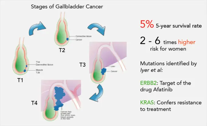
- Stage I. At this point, gallbladder cancer is restricted to the internal layers of the gallbladder.
- Stage II. This stage of gallbladder cancer has developed to affect the external layer of the gallbladder.
- Stage III. At this stage, gallbladder cancer has developed to affect one or more nearby organs, which can be the Stomach, Liver, or small intestine. The gallbladder cancer may have circulated to nearby lymph nodes also.
- Stage IV. The latest phase of gallbladder cancer comprises of large tumors that affect multiple close organs and tumors of any size that have spread to distant areas of the body.
Treatment
What gallbladder cancer treatment choices are available will rely on the stage of cancer, one’s overall health, and preferences.
The initial objective of treatment is to remove the gallbladder cancer, but when that isn’t possible, other therapies may help regulate the spread of the disease and keep a patient as comfortable as possible.
Surgery for early-stage gallbladder cancer
Surgery may be an alternative if one still have early-stage gallbladder cancer. Other choices include:
Early gallbladder cancer that is restricted to the gallbladder can be treated with an operation to remove the gallbladder, which is also called a cholecystectomy.
Gallbladder cancer that has expanded beyond the gallbladder and into the liver can be treated with surgery to extract the gallbladder, as well as parts of the liver and bile ducts that surround the gallbladder.
It’s not apparent whether extra treatments after successful surgery can improve the odds that the gallbladder cancer won’t return. Some researches have established this to be the case, so in some examples, the doctor may suggest radiation therapy, chemotherapy, or both after surgery.
So a patient may discuss the possible advantages and dangers of additional treatment with the doctor to discern what’s right.
Treatments for late-stage gallbladder cancer
Surgery can’t treat gallbladder cancer that has to disperse to other parts of the body. Rather doctors use procedures that may ameliorate the signs and symptoms of cancer and make the patient as comfortable as possible.
Options may include:
- Chemotherapy which is is a drug therapy that uses chemicals to eliminate cancer cells.
- Radiation is also an option that utilizes high-powered beams of energy, such as X-rays and protons, to destroy cancer cells.
There are such Clinical trials that are studies testing experimental or new medications to treat gallbladder cancer. Consultations should be held with the doctor to know whether one is qualified to partake in a clinical trial.
Procedures to relieve blocked bile ducts
Advanced gallbladder cancer can result in blockages in the bile ducts, resulting in further difficulties. Procedures to reduce blockages may benefit the patient. For example, surgeons can position a hollow metal tube (stent) in a duct to keep it open or surgically bypass bile ducts around the blockage.


