

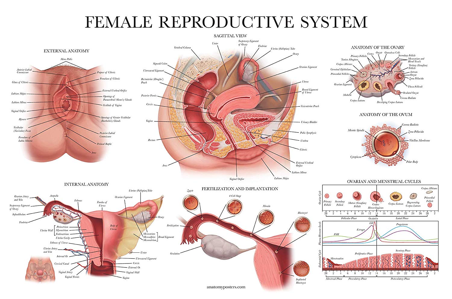
The components of the female reproductive system sustains and produce the female egg cells or ova and transfer these cells to a site where they can be fertilised by sperm; providing an adequate environment for the development of the fetus and also moves the fetus, outside the body, at the end of the gestation period, and provides the female sex hormones.
The female reproductive system consists of the external and internal sex organs that are responsible for the reproduction of offspring.
In human beings, this system is usually premature at birth and develops steadily to maturity at puberty to produce the gametes and to carry a fetus to a full period of gestation.
The internal sex organs are the Fallopian tubes, ovaries, and the uterus or womb, which houses the embryo during pregnancy. It also produces uterine and vaginal secretions, which aid the movement of sperm to the Fallopian tubes, while the ovaries provide egg cells.
The external sex organs are known as the genitals and composed of the libias, clitoris, vaginal opening, and the vulva.
The female reproductive cycle is all the processes that involve the production of egg cells or ovum and also the preparation of the uterus to accept a fertilised ovum to begin pregnancy.
If an ovum is provided but not fertilised and implanted to the walls of the uterus, it results in menstruation. The reproductive cycle takes about 28 days, but can be as brief as 24 days or even as long as 36 days for some women.
The stages of the female reproductive cycle
Oogenesis and Ovulation
The follicle-stimulating hormone (FSH), and luteinising hormone (LH) influences the ovaries to produce a fully developed ovum in a process known as ovulation.
Fourteen days into the reproductive cycle, an oocyte attains maturity and is expelled as an ovum. The ovaries usually develop many oocytes each month, but only one of them is released as an ovum.
Menstruation
While the mature ovum and moves through the fallopian tube, the endometrium develops and grows in preparation for the development of the embryo.
If the ovum is not fertilised or fails to be introduced to the endometrium, arteries in the uterus contracts and squeeze to cut off the flow of blood to the endometrium, this lack of blood flow spurs cell death in the endometrium and results in the shedding of tissue in a process called menstruation; this shedding begins around day 28 and goes on into the first days of the new reproductive cycle.
Fertilisation
Once the mature ovum is discharged from the ovary, the fimbriae trap the egg and take it down the fallopian tube to the uterus. It takes about seven days(a week) for the ovum to traverse to the uterus, if it meets with sperm, a single sperm can penetrate and fuse with the egg or ovum, fertilising it into a zygote.
A zygote is a coalition of the sperm cell and the egg cell. The zygote starts up as a single cell but eventually divides quickly; After 14days duration of cell division, the zygote becomes an embryo, and the embryo becomes a fetus.
The Formation of Zygote
For reproduction to happen, a single sperm cell must permeate the outer layer of an ovum. In most cases, an egg cell is released during the ovulation period of a woman’s monthly reproductive cycle.
Generally, thousands of spermatozoa venture to enter this single egg cell during sexual intercourse. So when one sperm enters the egg cell, chemicals in the surface layer of the egg cell stops other sperm cells from entering, and this other sperm cells eventually die.
Fertilisation occurs in the Fallopian tubes and commemorates the start of embryogenesis.
This process usually occurs as a result of sexual intercourse between the male and the female nevertheless, medically enhanced fertilisation is also feasible. The two conventional medically enhanced methods are: Intrauterine insemination (IUI) and in vitro fertilisation (IVF)
During an IUI, spermatic fluid is deposited into the uterus with the aid of a catheter so that the fertilisation can take place inside the woman’s body while in IVF, eggs are withdrawn from the ovaries and fertilised in a laboratory, and the zygote is then implanted into the body of the woman.
After fertilisation, the zygote splits through a process called mitosis, in which each cell duplicates by splitting up into two cells. This stage, which happens in two-weeks, is called the germinal period of growth and spans through the time of conception to implantation of the embryo.
In most cases, both the female and male sex cell contains 23 chromosomes. The egg cell contains genetic material from the mother, while the sperm cell contains genetic material from the father. Because each cell carries half of the genetic material, the cell is called the haploid cell.
When these two haploid cells come together, they become a single diploid cell that comprises of a total of 46 chromosomes. The zygote then moves down the fallopian tube to the uterus, where it implants itself to the lining of the uterus to obtain the nourishment it needs to survive.
The zygote stage lasts about four days. After which the mass of cells becomes known as a blastocyst on the fifth day of the germinal phase, this phase will last approximately 2weeks, after which the embryonic stage will commence.
The second stage lasts from two weeks after conception to the eighth week, where the organism becomes an embryo. In the ninth week after conception, it advances to the fetal stage, and from this point until birth, the organism becomes known as a fetus.
Pregnancy
When the ovum is fertilised by a sperm cell, the fertilised embryo usually implants itself to the uterine wall and forms a placenta, umbilical cord, and even an amniotic cavity. This embryo matures steadily until birth in a process called pregnancy.
Lactation
Lactation is the process of milk production and release of this produced milk to feed an infant; his process begins before birth controlled by prolactin a hormone.
After delivery, milk is released under the influence of a hormone called oxytocin, whose secretion is stimulated by the suckling of the nipple by the baby.
Milk is produced as long as breastfeeding takes place, but eventually, when the infant is weaned, milk production ends.
The release of milk by the nipples is a process called the “milk-letdown reflex” and is controlled by the oxytocin, which is dispatched in response to the infant’s suckling. Hence milk is released only when an infant is actively feeding.
Anatomy of the female reproductive organ
Ovaries
The ovaries are a ductless pair of glands with the shape and size of an almond; they are a primary female reproductive organ(gonad).
ovaries release eggs for possible fertilisation, they produce female sex hormones such as progesterone and oestrogen, and they protect the eggs a female is born with; All-female humans are born with two ovaries which stem from the uterus.
The ovaries reside on both sides of the uterus cushioning against the pelvic wall in a region known as the ovarian fossa, as they are firmly held in place by ligaments affixed to the uterus.
The primary function of the ovaries is to protect and shelter the eggs as women are born with their lifetime allotment of eggs
Secondly, the ovaries produce reproductive hormones called estrogen and progesterone, and the relaxin and inhibin. There are three varied types of estrogen: estradiol, estriol, and estrone.
They are employed by the body to develop the adult female characteristics, such as larger hips, breasts, etc. and to help in the reproduction cycle while progesterone aids reproduction. Relaxin helps the pelvic ligaments stretch during labour. Inhibin deters the pituitary gland from producing hormones which
Thirdly, ovaries release one egg each menstrual cycle in a process called ovulation. Within each ovary, there exist follicles, and at the core of each follicle is an inactive egg.
When a female child is reproduced, she carries 150,000 to 500,000 follicles in her premature ovaries. By the time she attains sexual maturity, the female has about 34,000 follicles.
When an egg is accelerated to maturity by hormones released from the pituitary gland, the follicle moves to the wall of the ovary. Where, the egg and the follicle matures and, when developed, become ready for ovulation. Mature follicles, also known as the Graafian follicles, can grow up to about 1.2 inches.
The follicle with the mature egg breaks, releasing the egg into the closest fallopian tube. From there, the egg drifts to the uterus. The body secretes the progesterone hormone thickens the lining of the uterus to receive the incoming egg.
This hormone is created by new cells growing where the old egg once inhabited in the ovary. These cells act as temporal glands and are called the corpus luteum.
The ovaries can be affected by cysts like polycystic ovary syndrome( PCOS) and cancer such as ovarian cancer. These disorders can lead to infertility and other high-risk diseases that can cause death.
Uterus
The uterus, also called the womb is a muscular, hollow, pear-shaped organ located between the urinary bladder and the rectum; it surrounds and provides support for the developing fetus during pregnancy.
The inner walls of the uterus, called the endometrium, provides assistance to the embryo during early development. The visceral muscles of the uterus compress during childbirth to propel the fetus out of the body, through the birth canal.
The uterus has four major areas that include;
- The fundus is the broad curved upper area where the fallopian tubes connect to the uterus.
- The body of the uterus, which is the central part of the uterus, begins directly below the fallopian tubes and continues below til the uterus walls and cavity narrows.
- The isthmus is the narrow, lower neck region, which is also the lowest.
- The cervix continues downward from the isthmus until it empties into the vagina.
The uterus generally is 2.4 to 3.1 inches long; its wall thickness is estimated to be around 0.8 to 1.2 inches. The width of the organ differs but is typically about 2.4 inches wide around fundus and only 1.2 this distance at the isthmus. The cavity in the uterus opens into the vaginal cavity, which makes up the birth canal.
The lining of the walls of the uterus is a moist mucosal membrane called the endometrium. The lining shifts in density during the menstrual cycle, and usually thickest during the release of eggs from the ovaries when the egg is fertilised, it hinges itself to the dense endometrial wall of the uterus and starts its development, but if unfertilised, the endometrial wall secrets its outer layer of cells with the eggs. They are passed from the body during menstruation.
The endometrium also keeps the egg and the sperm cells alive by secreting some substances. The constituents of the endometrial fluid include proteins, glucose, sodium, water, potassium, iron, and even chloride.
Proteins help in the implantation of the fertilised egg cells; glucose nourishes the reproductive cells while the other constituent provide a sustainable environment for the sperm and egg cells
The uterine wall is composed of three layers of muscle fibres. The muscle tissues run obliquely, longitudinally, and circularly, interwoven between connective tissues of, fibres which are elastic, collagen fibres, and blood vessels.
This strong muscular wall and becomes thinner as a result of expansion as a child grows inside the uterus. Subsequently, after birth, the uterus goes back to its original size in about eight weeks.
Vulva
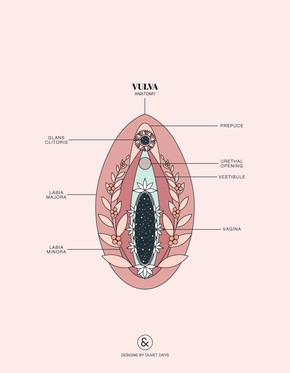
The vulva is the combined name all the external female genitalia found in the pubic region of the female body.
The vulva encircles the outer surface of the urethra and the vagina and includes the labia minora, labia majora, clitoris, and mon pubis. The mons pubis is an elevated layer of adipose tissue in between the pubic bone (that cushions the vulva) and the skin.
The mons pubis which it divides into two halves called the labia majora and are both usually covered up with pubic hairs.
Residing inside the labia majora is another hairless and smaller folds of skin called the labia minora, which surrounds the urethra and vaginal openings. On the other end of the labia minora is an erectile tissue known as the clitoris that has many nerve endings for sensing sexual pleasure.
Fallopian Tubes
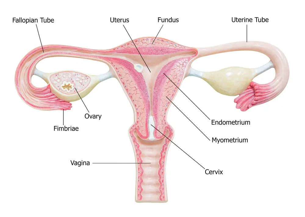
The fallopian tubes are a pair of muscular tubes that connects from the left and right corners of the uterus to the edge of the ovaries with delicate hair-like structures.
These hairs-like structures help to transport eggs to travel from the ovaries to the uterus.
The fallopian tubes end in a funnel-shaped, which ends with a small finger-like structure called fimbriae.
A blocked fallopian tube can cause infertility and usually shows no symptoms except inability or difficulty to conceive. This medical condition is called tubal occlusion, which can be caused by the previous history of pelvic infections, abdominal surgery, a burst appendix, endometriosis, Sexually transmitted diseases, etc.
Vagina
The vagina receives the penis during coitus. It is a muscular and elastic canal with a flexible, soft lining that aids lubrication and sensation.
The vagina opens the uterus to the outside world; the vulva forms the vestibule of the vagina. The vagina serves as a duct or channel for menstrual flow from the womb, and also serves as the birth canal where the baby passes during childbirth.
It has the hymen, which is a thin membrane of tissue tapers, and covers the vaginal opening. It can be ruptured by physical activities like exercise and sexual activities.
Below are some disorders of the vagina;
- Vaginitis
- Vaginismus
- Chlamydia
- Gonorrhoea
- Genital warts
- Trichomoniasis
- Bacterial vaginosis
- Vaginal cancer
- Vaginal prolapse
- Herpes
- Etc…
Breasts and Mammary Glands
The breasts are organs of the female body that contains milk ducts, adipose tissue, and mammary glands.
The two breasts are located on the left and right sides of the thoracic region or upper ventral region of the body. In the middle or hub of each breast is a nipple, which is highly pigmented and releases milk when stimulated.
The nipples are surrounded by a pigmented skin called the areola, which is responsible for protecting the tissues during breastfeeding. The mammary glands are a specific type of sudoriferous glands clustered within each breast that have been modified to produce milk to feed infants.
Fifteen or more clusters of mammary glands become activated during gestation and remain active until the infant is weaned. The milk departs through the milk ducts on its way to the nipple, where it leaves the body.
The effects of ageing on the female reproductive system.
When menopause happens, changes in the genital organs occur, and these changes can be very noticeable. The ovaries discontinue estrogen production, and the monthly menstruation stops.
After menopause, the tissues of the clitoris, vagina, urethra and Libia minora becomes thin, which can result in infections, vaginal discharges, dryness, and even chronic irritation. Also, the ovaries, uterus and fallopian tubes, become smaller.
With ageing, there is a reduction in the proportion of connective tissues in the ligaments, rectum, vagina, uterus, bladder and in muscles.
Consequently, the affected organs might prolapse, which can cause an impression of pelvic pressure or fullness, pain during coitus, loss of control over urination or bowel movement and difficulty urinating, the above problems are highly prevalent amongst women who have had many children.
The breasts decrease in size also as a result of the decrease in the production of estrogen, and the connective tissue that supports the breasts also diminishes, leading to sagging, which causes a change in shape. The breasts become less firm because of the replacement of fibrous tissues in the breasts with fat tissue.
These age-related changes do not diminish the sexual appetite of women. Some studies have shown that some women enjoy sexual activities better after menopause, possibly because the risks of getting pregnant is sufficiently reduced.
Besides, after menopause, the adrenal glands and ovaries continue to secrete male sex hormones, which helps maintain the slow the loss of muscle tissue, contribute to the overall well-being and increases sex drive.


")


