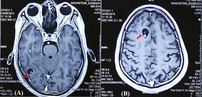
Cysticercosis also called Taeniasis is an infection where tapeworm larvae move out of the intestine and attack other organs like the brain, eye, or heart.
Cysticercosis is becoming an increasingly prevalent medical problem in the United States, especially in the Southwest and other areas where vast populations migrated from endemic regions, and populations often travel to these areas.
Cysticercosis is an uncommon infectious disease caused by the accumulation of the larval cysts of a tapeworm (cestode) within parts of the body.
The scientific name for the tapeworm that can cause cysticercosis is Taenia solium (T. solium), also known as the pork tapeworm. T. solium cysts (cysticerci) can affect any part of the body, including the brain, a condition called neurocysticercosis. Symptoms can vary from person to person.
If cysticerci are discovered in the brain, central nervous system anomalies may occur, often resulting in seizures and headaches. Cysticercosis can also affect the eyes, spinal cord, skin, and heart.
Causes of Cysticercosis
Cysticercosis can result from the ingestion of the eggs of the tapeworm known as Taenia solium. Ingestion of infected pork usually causes adult tapeworm infection, not cysticercosis.
When pig ingests tapeworm eggs, the move to the intestine and in the pig’s intestine, the eggs hatch and enter through the gut wall into muscle tissue where they develop into larval cysts known as cysticerci when the pig is killed, and the pork is consumed by a person, the cysticerci can enter and affix themselves to the intestine wall, where they develop into egg-producing adult tapeworms. This is called adult tapeworm infection and usually provokes no symptoms.
Nonetheless, individuals with adult tapeworm infection can get cysticercosis because adult tapeworm will release T. solium eggs through their feces. People can potentially ingest eggs through poor hygiene (autoinfection). These individuals may also regurgitate T. solium eggs from the intestines into the stomach.
Cysticercosis usually results when people eat food, especially pork, contaminated with T. solim eggs (rather than the larvae). The eggs travel through the bloodstream, ultimately finding their way into the muscle, subcutaneous, brain, and other tissues.
After 60 to 90 days, the eggs become enclosed in a cyst and form larval cysts (cysticerci). The cysts stay in the body tissue indefinitely, unable to continue to the next stage of their life cycle.
As long as these larvae remain alive, they appear to “disguise” themselves from the host’s immune system causing only minor symptoms. However, finally, the larvae die off, provoking a strong immune defensive response against it or the cyst enclosing it.
The cyst itself can become massive. Such inflammatory reactions can result in severe illness, particularly if the cysticerci are inhabited in the central nervous system or heart.
Risk Factors
The disease’s main risk factors are close association with pigs and drinking water or eating food infected with tapeworm eggs from porcine and even human feces.
Poor hygiene conditions can cause autoinfection. Persons living with a person who has a tapeworm infection are at increased risk.
Cysticercosis is not deemed contagious, but if infected people make poor sanitary choices (for instance, not washing hands after passing stool), they can infect others if the person accidentally ingests a parasite egg.
Rinsing and peeling all raw vegetables and fruits before eating can prevent transmission. The incubation period is estimated to be three and a half years, but it can also have ten days to 10 years.
Symptoms
The symptoms of cysticercosis differ from person to person, depending upon the amount and location of cysticerci within the body. Cysticerci can be discovered in the muscle tissue.
In some instances, the cysts have been located in the brain, eyes, or heart tissue. Some individuals with cysticercosis will demonstrate they are asymptomatic or display mild symptoms.
Many persons with cysticercosis have central nervous system involvement (neurocysticercosis). However, many individuals with neurocysticercosis do not display or are usually asymptomatic.
The exact symptoms of neurocysticercosis depend upon the number and location of cysts involved and an individual’s immune system reaction. The four basic forms of neurocysticercosis are parenchymal, subarachnoid, intraventricular, and spinal.
Symptoms prevalent to all types of neurocysticercosis include seizures and accumulation of excessive cerebrospinal fluid (CSF) in the skull (hydrocephalus), which results in increased pressure on the brain’s tissues resulting in a variety of symptoms including dizziness, headaches, nausea, changes in vision, and vomiting.
In some cases, people who develop hydrocephalus often, in turn, develop swelling of the optic disc (papilledema). Papilledema may result in blurred or double vision.
The parenchymal disease may be linked with behavioral changes, intellectual impairment, headaches, seizures, hydrocephalus, muscular weakness on one side of the body (hemiparesis), and the ability to coordinate voluntary movements (ataxia) can also occur with this type of neurocysticercosis.
Subarachnoid cysticercosis can be associated with severe Inflammation of meninges (the membrane covering the brain), hydrocephalus headaches, and seizures. Intraventricular cysticercosis may result in obstructive hydrocephalus.
A form of this kind of cysticercosis known as racemose cysticercosis may occur. Racemose cysticercosis is characterized by the accumulation of cysts at the base of the brain, potentially resulting in mental deterioration, coma, and life-threatening complications.
Cysts affecting the spinal cord are usual but can result in Inflammation of the brain and spinal cord membranes, or compression of the spinal cord.
In some cases, individuals may experience heavy central nervous system infections, potentially causing life-threatening complications such as stroke or coma (cysticercal encephalitis). Individuals with heavy CNS infections often develop muscle pain (myalgia), weakness, and fever.
Ocular cysticercosis can develop when cysts form in the eyes. Symptoms linked with this can include pains in the eye, loss of vision, and separation of the retina (nerve-rich membrane lining the eyes) from retinal detachment (underlying supporting tissue). In some instances, cysticercosis can affect the eyes (isolated ocular cysticercosis).
In some instances, cysts may appear under the skin resulting in small lumps. These lumps usually do not cause any extra symptoms.
Since a sudden onset seizure is often the indicating sign, the first doctors to diagnose the disease are emergency doctors when they conduct a CT scan on the head. Doctors who can also treat and manage the patient are neurosurgeon, neurologist, or an infectious disease specialist.
Diagnosis of Cysticercosis
The diagnosis of cysticercosis is usually founded on clinical presentation, abnormal findings on neuroimaging and serology (blood test like an immunoblot assay), and sometimes a biopsy.
Conditions for diagnosis include absolute criteria (histologic demonstration of the parasite, direct visualization of subretinal parasites and cystic lesions displaying the scolex, the anterior end of a tapeworm with suckers and hooks for attachment).
Medical professionals may use additional major and minor criteria if absolute criteria are not met.
Home Remedies for Cysticercosis
There are no home remedies for cysticercosis. However, there are believes that papaya, pineapple, garlic, cloves, and pumpkin seeds preparations can relieve a person of tapeworms.
One should discuss such methods with the healthcare provider, especially if one is pregnant, before using these methods.
Treatment for Cysticercosis
Most patients with cysticercosis usually experience no symptoms or evidence to indicate cysticercosis, so antiparasitic therapy can be beneficial to them.
Nonetheless, in symptomatic patients, medical and surgical treatments are obtainable. For instance, physicians can treat ocular cysticercosis with albendazole, corticosteroids, and surgery to remove the cysticerci.
A doctor may surgically remove an inflamed granuloma in the muscle caused by a cysticercus, while cerebrospinal fluid may need surgical diversion if hydrocephalus develops.
Some persons with neurocysticercosis may be treated with albendazole or praziquantel, corticosteroids, and antiseizure medications. The doctors will help select the appropriate treatments for each individual.
Complications of Cysticercosis
The disease’s complications may include paralysis, hydrocephalus, chronic meningitis, vasculitis, partial blindness, seizures, coma, brain edema, and even death. For instance, an 18-year-old male in India went to the ER because of seizures.
His head MRI showed a substantial number of cysts in the brain. He died two weeks later.
Prognosis of Cysticercosis
The prognosis for cysticercosis is good for 80% or more who experience no symptoms. The prognosis begins to become worse as complications increase.
Prevention of Cysticercosis
It is possible to prevent the disease by preventing people from swallowing eggs from the parasites. This can be accomplished by limiting the contamination of food and water from pig and human feces.
Eliminating undercooked pork from the diet reduces intestinal infection rates by tapeworms. Washing and peeling all raw vegetables and fruits before eating can help prevent transmission.
Reference;


