

A computerised tomography (CAT or CT) scan is an imaging technique that provides a detailed cross-section image of the body. It makes use of rotating x-ray machines and computers to create these images.
These images are more detailed than normal x-ray results. They can show the blood vessels, bones and soft tissues in different parts of the body. A CT scan is useful for collecting images of
- Brain
- Chest
- Heart
- Spine
- Shoulders
- Abdomen
- Knees
- Soft tissues
- Lungs
- Blood vessels
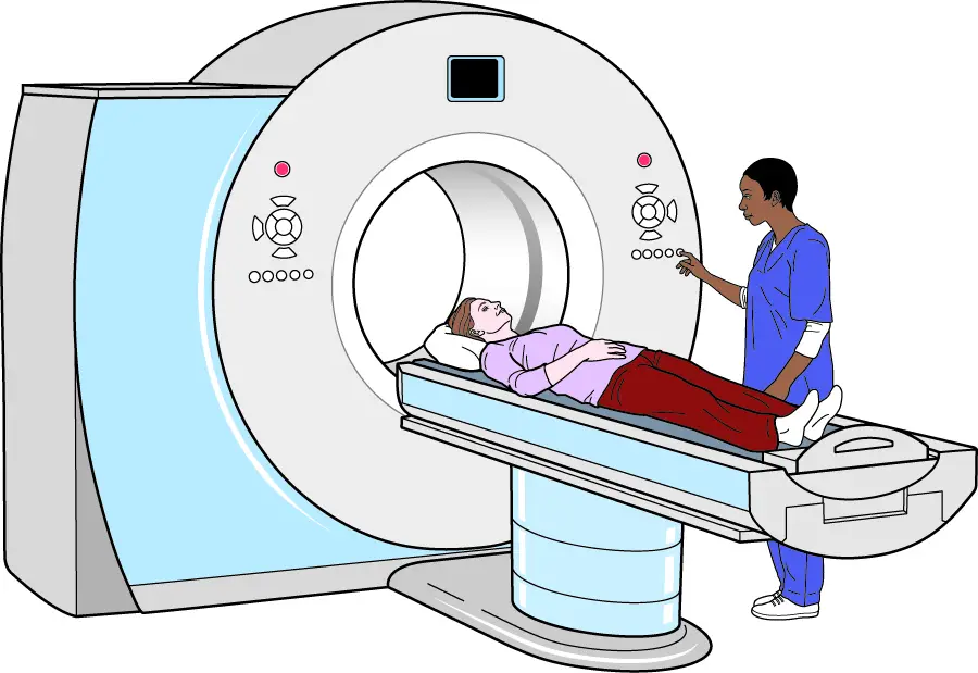
During a CT scan, the patient lies down on a motorised examination table that slides into a tunnel-like machine.
Once inside, the scanner rotates and takes a series of x-ray images from different angles.
The resulting images are then sent to a computer where they’ll be combined to create pictures of cross-sections or slices of the body. They can also be merged to form a 3D image of a particular part of the body.
Why is a CT scan performed?
This imaging technique has many uses, but, it is specifically well-suited for evaluating injuries and diagnosing diseases. A CT scan can help your doctor
- Diagnose bone fractures, infections and muscle disorders.
- Examine blood vessels and other internal organs and structures.
- Discover the location of masses and tumours (including cancer)
- Examine the extent of internal bleeding and internal injuries
- Guide procedures such as biopsies and surgeries
- Track the effectiveness of treatments for certain diseases, including heart disease and cancer.
The procedure is minimally invasive and can be carried out quickly.
How is a CT scan done?
Preparing for the scan
Before the scan, patients are advised to abstain from food or possibly drink for a few hours – maybe four or five hours – especially if your doctor is planning on using a contrast material or agent.
Contrast agents are specially formulated dye that helps highlight the body parts being examined.
The contrast agent blocks x-rays and appears white on the scan, allowing it to highlight blood vessels, intestines or other structures.
Contrast agents are usually made of Barum sulfate or iodine. Depending on the area being examined, your doctor may administer the contrast in one or more of three ways
- Orally: you may be asked to drink a liquid containing the contrast material. This method helps illuminate your oesophagus or stomach. The drink may taste unpleasant.
- Injection: alternatively, the contrast material can be injected into a vein in your arm. This will help your blood vessels, urinary tract, gallbladder, or liver to stand out during imaging. You may feel a metallic taste in your mouth or warmth in your vein during the injection.
- By enema: if your intestines are being scanned, the contrast agent may be inserted into your rectum. You may feel bloated and uncomfortable during this process.
Before the scan, you may be asked to change into a hospital gown and remove any jewellery or clothing with metal buttons. Jewellery and metallic objects (including dentures) can distort the visualisation process.
During the scan
After that, you’ll be asked to lie on a table that slides into the scanner. You doctor will exit the exam room and go to a control centre where he can monitor the whole visualisation process. You’ll be able to communicate with him via intercom.
As the table slides into the scanner, x-ray tube and detectors slowly rotate around you and produce an image slice of your body. After one image slice, the table tilts a bit for the machine to take another shot, and so on. You may hear buzzing, whirring and clicking noises during the scan.
The patient may need to lie very still for the best results. Pillows and straps may be used to secure the patient to the table and help reduce movement.
Your doctor may tell you to hold your breath for a while during the scan to reduce the movement of your chest.
Your doctor may recommend a sedative for little children who need a CT scan to keep them from moving. Most times, the patient lies on their back facing up. But in some cases, they may need to lie sideways or face down.
The entire visualisation process may take anywhere from 20 minutes to an hour.
After the scan
At the end of the scan, the images are sent to a radiologist to examine them. A radiologist is a doctor who specialises in diagnosing and treating medical conditions using visualisation techniques such as x-rays and CT scans. The radiologist will then send the results to your doctor who’ll then explain them to you.
If you were given a contrast agent before the scan, the doctor would advise you to drink a lot of fluids to help flush out the contrast materials from your system.
What are the health risks associated with a CT scan?
Risk of radiation
A CT scan uses a small dose of radiation during the process of visualisation. It may expose you to more radiation than standard x-rays. However, your chances of getting cancer from the radiation are very slim if you get just one scan.
Taking multiple x-rays and CT scans over time may increase your risk of cancer. Children receiving CT scan are at a higher risk of cancer, especially when taking the radiation to the chest and abdomen.
Allergic reaction
Most contrast materials contain iodine which may have some side effects on those who are allergic to iodine. Be sure to notify your doctor about any previous reaction to iodine.
If you must be given a contrast agent, your doctor may give you steroids or allergy medications to counteract any potential side effects.
Pregnancy
Also, you must inform your doctor if you’re pregnant. Though it is unlikely that the radiation from the CT scan will be harmful to your baby, your doctor may suggest another exam such as an MRI scan or an ultrasound, to minimise the risk.
What do CT scan results mean?
Results from the exam are considered to be normal if the radiologist didn’t spot any abnormality in the images such as blood clot, tumours or fractures.
If the radiologist discovers any abnormality in the results, he may recommend further tests and treatment, depending on the type and severity of the abnormality.


")


